What is Acquired Digital Fibrokeratoma?
Acquired digital fibrokeratoma (ADFK) is a relatively rare, non-dangerous growth made up of fibrous tissue. Like the name suggests, it was first reported in 1968 when multiple occurrences were observed mainly on fingers. While it’s most common on fingers, it can also appear on other extremities like heels, toes, palms, and the bottom of the foot, which is why it sometimes gets referred to as an acral fibrokeratoma.
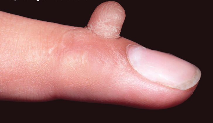
You’d typically identify ADFK as a small, solitary bump surrounded by a thickened ring of skin. Doctors usually consider possibilities like an extra, undeveloped finger or a common wart when trying to diagnose this condition. The recommended treatment is surgical removal of the growth, because unlike some conditions, ADFK doesn’t go away on its own.
What Causes Acquired Digital Fibrokeratoma?
Doctors aren’t entirely sure what causes acquired digital fibrokeratoma, a skin condition often found on hands and feet. There’s a theory that it might develop due to an injury or constant irritation, especially in areas like the hands and feet that are often exposed to injuries.
A look under the microscope shows that there are a lot of tightly packed collagen fibers in the areas affected by this condition. This suggests that new cells that make collagen might be involved in causing the disease.
However, most patients don’t recall having any specific injury at the spot where the skin condition shows up. This has led to two other theories about what might cause this condition.
One theory suggests an infection could be a potential cause. There have been a couple of cases where this condition developed after an infection caused by a type of bacteria called Staphylococcus aureus. This might indicate that an infection could create the same results under a microscope as an injury would when it comes to the development of this condition.
Another theory is that this skin condition could be due to a benign tumor. There is a certain evidence that supports this idea. There was a recorded case where this condition was observed alongside an overgrowth of gum tissue after a certain treatment.
Risk Factors and Frequency for Acquired Digital Fibrokeratoma
Acquired digital fibrokeratoma is a rare type of tumor, and we’re still not sure how often it occurs. Most of what we know comes from individual case reports, and these often focus on how unusually big the tumors are, where they are found, or what other conditions they’re associated with. However, there have been a few series of cases published, with the largest one including 50 patients.
- Acquired digital fibrokeratoma has been seen in patients from all different racial backgrounds.
- Cases have been reported in several languages, including German, Polish, Japanese, French, and Indian.
- It seems to affect males more often than females.
- It’s been reported in people from ages 12 to 70, but is most likely to occur in middle-aged adults.
Signs and Symptoms of Acquired Digital Fibrokeratoma
Acquired digital fibrokeratoma is a condition that typically doesn’t present any symptoms but usually appears as a small bump on the skin. This bump is usually skin-colored, well-defined, and often has a distinctive thickened ring at the base. Most commonly, they appear on fingers and toes, but they can also show up on the lower lip, nose, elbow, knee, or around the nails. These bumps are usually smaller than 1 cm, but there have been cases where they are larger, and these are known as ‘giant’ acquired digital fibrokeratomas.
There is a specific form of this condition known as multibranched acquired periungual fibrokeratoma (MAPFK). In cases of MAPFK, there are several thread-like bumps along the cuticle that look similar to warts caused by the human papillomavirus (HPV).
Dermoscopy, a technique of looking at the skin using a tool that magnifies the area, can be used to examine these lesions. Dermoscopy has revealed thread-like structures and varied findings due to potential impacts on blood vessels and the build-up of collagen fibers within the lesions.
Testing for Acquired Digital Fibrokeratoma
The skin-related characteristics of a condition called acquired digital fibrokeratoma haven’t been widely studied, and might vary among cases. Researchers Rubegni and colleagues noted a consistent pale-yellow core surrounded by a white, flaky ring. These details may signify thickened skin, hardened skin, and a concentration of dense, tough tissue. A white-yellow area with tiny blood vessels was noticed around the edges, while rounded blood vessels surrounded the whole tumor.
In another study by Hayashi and colleagues, red-clustered structures separated by white crisscrossed skin sections were reported. These observations could reflect the pulling back of the skin layer according to the microscopic tissue analysis.
Treatment Options for Acquired Digital Fibrokeratoma
Acquired digital fibrokeratoma (ADFK) can be treated in several ways including cryotherapy, shave excision, curettage and cauterization. However, the most effective treatment is generally considered to be surgical removal of the tumor. This is primarily to avoid the risk of the tumor coming back.
When the fibrokeratoma is found on the nail plate, sometimes the whole nail or part of it may need to be removed or another form of surgery may be needed. In these situations, you may need a specialist such as a dermatologist or a surgeon specializing in Mohs micrographic surgery who is skilled in nail surgeries.
There have been cases where ADFK is linked to a variety of conditions and factors. An infection from Staphylococcus aureus bacteria has been involved in some cases. It has also been associated with leprosy, especially in people who have diminished sensation.
ADFK has also been linked with conditions like brain infarction, use of cyclosporine in kidney transplant recipients, skin growth called pyogenic granulomas, and certain skin conditions like pityriasis amiantacea, and psoriasis vulgaris. However, the reasons for these connections are not fully understood and may be due to skin picking behavior causing trauma to the affected areas.
With its similar microscopic features to another condition known as Koenen tumor, there has been speculation about a potential link between ADFK and tuberous sclerosis, a genetic disease that causes growths in various parts of the body. While there are no firm guidelines about this, it is generally recommended that individuals with multiple ADFK may need to be carefully checked for other signs of tuberous sclerosis.
What else can Acquired Digital Fibrokeratoma be?
Recognizing skin growths that look like acquired digital fibrokeratoma (ADFK) is important for proper and timely treatment. There are several conditions that can resemble ADFK, including:
- Supernumerary digit: This is an inherited bodily abnormality usually found at the base of the pinky finger. It’s different from ADFK because it involves nerve bundles and neuroid elements. However, it could be mistaken for ADFK, as reported in a case study where ADFK was diagnosed on the big toe.
- Verruca vulgaris or common warts: These are the most common growths on the hands and fingers caused by certain types of human papillomavirus (HPV). They usually have tiny black spots known as thrombosed capillaries. There have been cases where ADFK was incorrectly diagnosed as warts and it reappeared after treatment.
- Cutaneous horn: This is an outgrowth on the skin that contains keratin. It requires examination as there could be an underlying disease that might need treatment. Cutaneous neurofibromas, which are soft, dome-shaped tumors on the skin, can look like ADFK.
- Superficial acral fibromyxoma or digital fibromyxoma: These are lesions that could be mistaken for ADFK due to their similar appearance. They might show more cell presence in lab tests. Also, specific tests for protein markers CD34 or CD99 are positive while they are negative for ADFK.
- Koenen tumor: This is a growth that shares many characteristics with ADFK. However, Koenen tumor is associated with a genetic disease called tuberous sclerosis.
- Eccrine poroma: These are common types of skin buds that might appear on the hands and feet. They look different under the microscope because they show cube-shaped cell growth and intercellular bridges, which isn’t the case with ADFK.
- Pyogenic granuloma: This is a red, dome-shaped growth that usually occurs where there has been minor injury. Alongside this, other conditions like dermatofibroma, papillary digital adenocarcinoma, or keratoacanthoma could also have similar symptoms.
So, it’s crucial for a doctor to consider these conditions and conduct the necessary tests to make an accurate diagnosis.
What to expect with Acquired Digital Fibrokeratoma
Acquired digital fibrokeratoma is a harmless tumor that does not pose a risk of becoming cancerous. Its removal through surgery is often successful and carries a low risk of pain, bleeding, and infection.
After undergoing surgery, patients can look forward to a full recovery and a pleasing cosmetic result. With the right surgical treatment, the chances of the tumor returning are very low.
Possible Complications When Diagnosed with Acquired Digital Fibrokeratoma
ADFK, in most cases, does not result in severe complications. However, there’s a rare form known as periungual fibrokeratoma that might lead to changes in the shape of the nail and impact the functionality of the finger.
Preventing Acquired Digital Fibrokeratoma
The root cause of ADFK isn’t completely understood, which means there aren’t any known ways to prevent it. It remains crucial though, to educate patients on how to care for themselves after surgery. This can help lessen the chances of complications like infections.


