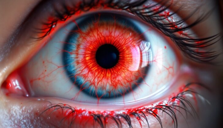
What is Cherry Red Spot?
The term “cherry-red spot” describes a red area in the middle of the macula. The macula is a part of the eye, and it’s surrounded by an area called retinal opacification, which may appear in certain health conditions. This retinal opacification could be caused by many factors including the build-up of various substances in certain eye cells (known as ganglion cells) during storage disorders, or due to insufficient blood supply or damage to the retinal tissue, like in the case of central retinal arterial blockage.
If a patient has a cherry-red spot at the macula, it’s very important to rule out serious diseases that could threaten their life or vision. The treatment approach for a patient with a cherry-red spot will vary depending on the root cause of the problem.
What Causes Cherry Red Spot?
A cherry-red spot can be caused by various health problems.
For instance, it could be a sign of a sudden blockage in the central retinal artery (CRAO) in elderly males or females. This usually happens when a clot breaks away from the walls of arteries in the neck or less commonly, from the heart or the main blood vessel in the body, called the aorta. The clot then travels to the eye, blocking blood flow and resulting in a cherry-red spot. Inflammation of the arteries like giant cell arteritis could also cause CRAO.
Other things that could lead to a cherry-red spot include bleeding or a mass in the space behind the eyeball, anesthetic injections, leukemia affecting the optic nerve, tearing of the optic nerve, or death of tissue in the macula, the part of the retina responsible for clear, detailed vision.
Certain diseases can also result in a cherry-red spot. These include Tay-Sachs disease, which is a fatal genetic disorder that typically starts in infancy and mostly affects children of Ashkenazi Jewish heritage. Another condition is Sandhoff disease, which shares similarities with Tay-Sachs disease and is caused by a certain genetic mutation.
Blunt trauma can result in a condition called Commotio retinae, which can sometimes lead to a cherry-red spot.
Niemann-Pick disease, which results from the body not being able to break down certain fats and cholesterol, leading to their accumulation in the organs, can also cause a cherry-red spot.
Other genetic disorders that can lead to a cherry-red spot include GM1 gangliosidosis type 1, sialidosis, Farber lipogranulomatosis, metachromatic leukodystrophy, and galactosialidosis. Being overly nourished can also cause a cherry-red spot, as can inflammation of the central part of the retina.
Additionally, exposure to certain toxic substances can cause a cherry-red spot. These include quinine (a medication used to treat malaria and certain heart conditions), carbon monoxide, dapsone (a medication often used for skin conditions), methanol (a type of alcohol that can be poisonous if swallowed or absorbed), or the antibiotics gentamicin and amikacin when they are injected directly into the eye.
Finally, while Gaucher’s disease can cause a buildup of certain fats in the cells of various organs, the presence of a cherry-red spot in this disease is uncertain.
Risk Factors and Frequency for Cherry Red Spot
A cherry-red spot at the back of the eye is not a frequent occurrence and the amount of people who get it can vary depending on what causes it. One of these causes, Tay-Sachs disease, is estimated to only affect one in every 320,000 newborns. However, the exact number of people who experience a blockage in the blood supply to the retina (the condition known as Central Retinal Arterial Occlusion), particularly a sudden blockage lasting less than 48 hours, is uncertain. One study suggests it might affect approximately 0.85 out of every 100,000 people each year, or about 1.13 out of every 10,000 outpatient visits.
Signs and Symptoms of Cherry Red Spot
Central retinal artery occlusion is a condition that often affects older people, and it usually results in a sudden loss of vision. Typically, the patient may have an abnormal pupil response in the affected eye. An examination reveals a whiteness in the back part of the eye, which obscures visibility of the choroidal vessels. The center of the macula retains its color, appearing as a cherry-red spot. Other signs might include a fragmented blood column in the retinal vessels, which may move slowly. There might be a clot in the central retinal artery or its branches visible. In the later stages, the retina may thin, pigmentation may change at the macula, and the optic nerve may show signs of damage.
In lipid storage disorders, signs of brain degeneration and enlargements of the internal organs can be seen. Early signs in the eye include a white ring-like area around the macula with a cherry-red spot. In the later stages, the inner layers of the retina thin, the cherry-red spot disappears, and the optic nerve shows signs of damage. It’s very important to consider family history and to examine other family members. If a patient has had a commotio retinae (retinal bruising), they should have a history of a blunt trauma. Signs of blunt trauma could include bleeding in the front part of the eye, inflammation of the middle layer of the eye, and signs of trauma damage. Inflamed retinas may have associated inflammation within the eye, vitreous inflammation, and retinal tissue death more visible on retina scans.
- Sudden loss of vision
- Symptoms often start in elderly people
- Signs of trauma or presence of related medical conditions
- Abnormal pupil response in the affected eye
- Whiteness in the back part of the eye
- ‘Cherry-red’ spot in the center of the macula
- Changes in blood flow within the retinal vessels
- Possibility of a clot in the central retinal artery
- In later stages, thinning of the retina, pigmentation changes, and optic nerve damage
Testing for Cherry Red Spot
When dealing with issues related to the inner surface of the eye (the fundus), your doctor might take a color photograph of it. This photo helps in understanding the details of your eye health. Besides, it also aids in explaining the disease to you, following the progression of the disease, and analyzing the response to treatment.
You might also undergo a test known as Fundus Fluorescein Angiography (FFA). This test shows blood circulation in the eye. If you have a blockage in the central retinal artery (CRAO), FFA might show a slowed-down dye progression, indicating reduced blood flow. There could also be an abrupt ending to the dye at the blockage area. If the blockage has cleared, or if it was only temporary, the test might even look normal. In some specific diseases like lipid storage disorders, FFA might reveal an area of dark or lesser fluorescence (glow).
Then another examination called Optical Coherence Tomography (OCT) can be performed, which provides an image of your eye’s macula (the region responsible for sharp, straight-ahead vision). If you have CRAO, it might show enhanced reflection of some layers of the retina and thickening of the retina. For lipid storage disorders, OCT may demonstrate increased reflectiveness in certain parts of the retina. If there was an injury to the retina, also known as commotio retinae, OCT usually does not show an increased retinal thickness but may show increased reflectiveness in other regions.
In a test known as Autofluorescence, signs of a disease known as sialidosis may be indicated by increased autofluorescence – a natural emission of light – around the central part of the retina (fovea). Conversely, in the case of CRAO, this test might show diminished autofluorescence in the eye areas where the retina appears whiter due to the disease.
In suspected cases of lipid storage disorder, your doctor will assess whether there’s an enzyme deficiency and whether other body systems are affected. If retinitis (inflammation of the retina) is a concern, you might receive tests for your immune system status, a neurological examination, and possibly a sampling of eye fluids to check for different infectious causes.
Treatment Options for Cherry Red Spot
Manageing the cherry-red spot in your eye depends on what’s causing it. For Central Retinal Artery Occlusion (CRAO), a condition that is essentially a “stroke” in your eye, the treatment options might include eye massage, withdrawing fluid from the front of the eye to lower eye pressure, using medications to lower eye pressure, oxygen therapy, and treatments that break up blood clots. Though these treatments might help, there isn’t clear proof yet that these approaches can actually improve your final vision.
For a condition known as Giant Cell Arteritis (GCA) which can also cause CRAO, medications known as steroids are typically used to prevent vision loss in the other eye.
In treating storage diseases, or conditions where harmful amounts of substances build up in the body, it often takes a team of different specialists. Some general advice for managing these conditions might include limiting certain substances in your diet, replacing certain enzymes (a type of protein that helps speed up chemical reactions in the body), and managing symptoms as they come up.
Treating retinitis, or inflammation of the retina, will be based on the specific details of your condition, what your doctor sees when they examine your eyes, and the results of any tests you may have had.
If you have commotio retinae or Berlin’s edema, conditions that are often the result of trauma or injury, your doctor will need to check for other signs of injury, such as inflammation, blood in the eye, iris damage, or retinal damage. Any eye injuries that are found will need to be treated, and you may need to use steroid medications. Most people with commotio retinae recover on their own over time.
What else can Cherry Red Spot be?
When a person has a condition known as a “cherry red spot,” there are several other medical conditions that doctors might consider as other possible diagnoses. These include:
- Macular hole
- Macular pseudohole
- Laser injury to the fovea
- Macular telangiectasia type 2
- Choroidal neovascular membrane
- Macular hemorrhage
What to expect with Cherry Red Spot
The future outcome or ‘prognosis’ of a condition known as a ‘cherry-red spot’ on the macula, the part of the eye responsible for central vision, can change depending on what’s causing it. The prognosis refers to the likelihood of recovery or recurrence of a disease.
Most cases of ‘commotion retinae’, a condition where the eye’s retina is temporarily damaged, usually get better over time and the patient’s vision generally improves. However, when the ‘cherry-red spot’ is caused by Central Retinal Artery Occlusion (CRAO) or damage to the macula, the patient’s vision outcome is usually poor. Patients with CRAO may even end up with a painful blind eye due to another condition called neovascular glaucoma, which is when new blood vessels form on the eye’s iris.
Patients diagnosed with ‘progressive outer retinal necrosis (PORN)’, a rare and serious eye infection, often lose their ability to perceive light, indicating complete vision loss. In a different situation, when a ‘cherry-red spot’ is a symptom of a rare genetic disorder called Tay Sachs disease, it is usually fatal by five years of age.
Possible Complications When Diagnosed with Cherry Red Spot
The potential problems associated with a cherry red spot at the back of the eye depend on what’s causing it. For example, an eye condition known as CRAO can lead to thinning and decay in the retina and optic disc over time. A condition called Commotio retinae sometimes leads to a disorder where dark pigment spots appear on the retina and the retina becomes thin. For those with a lipid storage disorder, the optic nerve may appear pale, and the retina could thin as the condition progresses. Sometimes, those with a damaging inflammation of the retina called necrotizing retinitis might suffer from a detached retina.
Potential Problems:
- Retinal and Optic Disc Thinning with CRAO
- Pigmentary Retinopathy and Retinal Thinning with Commotio Retinae
- Optic Nerve Pallor and Retinal Thinning with Lipid Storage Disorder
- Retinal Detachment with Necrotizing Retinitis
Preventing Cherry Red Spot
If you see cherry red spots at the back of your eye, it’s important to understand the cause, what the future may hold, and how it can be managed. Such spots could potentially threaten your life or vision. Therefore, it’s crucial that these are explained to you in great detail so that you can make an informed decision about your treatment.
Lipid storage disorders, which are genetic conditions where fats build up in your cells and tissues, often cause these red spots. If you have this condition, genetic counseling can be very helpful. This process can help you understand the disorder you have, the risks it presents to your health, what can be done to manage it, and how it could affect your family.
For a condition known as Central Retinal Artery Occlusion (CRAO), where a blockage in the arteries in your eye impacts your vision, it is really important to manage and control possible risk factors tied to your blood vessels; for instance, taking good care of your heart
.


