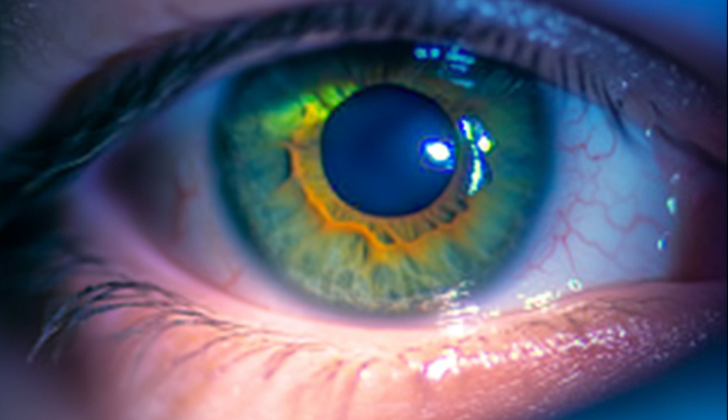
What is Neurotrophic Keratitis?
Neurotrophic keratitis is a type of eye disease that breaks down the cornea, the clear front surface of your eye, due to damaged sensory nerves in the cornea. This disease results in reduced or no feeling in the cornea, leading to the breakdown of the surface layer of the eye, difficulty in healing, and eventually leads to the development of corneal ulcers (sores), melting, and even rupture of the cornea. This condition was first identified as neuroparalytic keratitis – a condition that damages the cornea due to nerve paralysis, in a study conducted by Magendie in 1824.
What Causes Neurotrophic Keratitis?
Neurotrophic keratitis is a condition that can occur when there’s damage to the fifth cranial nerve, also known as the Trigeminal nerve. This nerve covers a vast area from the nerve center in your brain (Trigeminal nucleus) to the ends of the nerve in your eye (corneal nerve endings). Anything that impacts this nerve could lead to neurotrophic keratitis.
There are many potential causes of this damage. Common ones include an infection of the cornea caused by the herpes virus (herpetic keratitis), chemical burns in the eye, wearing contact lenses for a prolonged period, undergoing eye surgery or treatments for trigeminal neuralgia (a chronic pain condition affecting the Trigeminal nerve), and surgical treatments to fix a broken jaw.
Less common causes can include issues within the brain that take up space and put pressure on the nerve, like certain types of benign brain tumors (schwannoma, meningioma), or aneurysms. This pressure can decrease the sensitivity of the cornea. There are also certain health conditions that might affect the functionality of the Trigeminal nerve, such as diabetes, multiple sclerosis, and Leprosy, which can lead to this eye condition.
It’s rare for children to have neurotrophic keratitis, but when it does occur, it is often linked with congenital syndromes. These are disorders that a person is born with, such as Riley-Day syndrome, Goldenhar-Gorlin syndrome, Mobius syndrome, Familial corneal hypesthesia, and Congenital Insensitivity to Pain with Anhidrosis, a condition where someone can’t feel pain and can’t sweat normally.
Risk Factors and Frequency for Neurotrophic Keratitis
Neurotrophic keratitis is a rare eye disease, affecting less than 5 in every 10,000 people. It’s found in differing rates among various conditions:
- It is seen in 6% of people who have herpetic keratitis.
- It affects 12.8% of individuals with Herpes zoster keratitis.
- It occurs in 2.8% of patients who have had surgery for Trigeminal neuralgia.

Gaule spots. Gaule spots are scattered areas of dried epithelium, typically seen
in stage 1 neurotrophic keratitis.
Signs and Symptoms of Neurotrophic Keratitis
Neurotrophic keratitis is an eye condition that people often don’t notice symptoms for because of a lack of feeling in their cornea, which is the clear front surface of the eye. In some cases, people may notice eye redness and blurred vision which could be caused by surface wounds on the cornea, swelling, or scarring. This condition can be suggested by a history of eye pain and redness, skin blistering or scarring from previous herpes infection. Additionally, previous eye injury, surgery, chemical burns, long-term use of eye medication, nerve-related surgeries, or diabetes could also point towards having neurotrophic keratitis.
The main feature of neurotrophic keratitis is decreased or absent corneal sensation. The severity of corneal damage in Neurotrophic keratitis is classified into three stages, following the Mackie classification:
- Stage 1: Corneal epithelial changes with a dry and cloudy corneal exterior, punctate keratopathy (small, superficial wounds), and corneal edema (swelling).
- Stage 2: Frequent and/or lasting epithelial defects, usually seen at the top half of the cornea, which are oval or circular in shape.
- Stage 3: Corneal ulcer with deep corneal involvement potentially leading to corneal melting and corneal perforation.
In short, the stages of neurotrophic keratitis include epithelial changes (stage 1), lasting epithelial defects (stage 2), and corneal ulcer (stage 3).
Testing for Neurotrophic Keratitis
The diagnosis of a condition involving the trigeminal nerve, one of the primary nerves in the face, is often based on a person’s specific symptoms, coupled with a physical examination. This nerve is critical for eye health, so problems can lead to corneal issues like long-lasting wounds, ulcers, and decreased sensitivity.
Medical professionals typically take into account factors such as existing health conditions (like diabetes), use of certain medications, and possible eye-related causes (such as excessively wearing contact lenses or exposure to chemical burns). Testing other facial nerves can help pinpoint where the issue is. If there’s a problem with the seventh or eighth nerve, this could indicate a condition called acoustic neuroma, which could have been caused by damage to the trigeminal nerve either naturally or during a surgical procedure. Issues with the third, fourth, and sixth nerves could suggest a problem in a space within the skull known as the cavernous sinus.
Part of the evaluation involves checking the cornea with a special tool called a Cochet-Bonnet or no-contact gas esthesiometer, which helps measure how sensitive the cornea is. A device named the slit lamp can highlight the characteristic lesions on the cornea and indicate if the iris is deteriorating, which is typical of herpes infections. If an ulcer is found, it must be tested to rule out potential infections.
In some instances, a thorough eye examination may reveal swelling or paleness of the optic disc, the point of exit for nerve fibers from the eye to the brain, indicative of an intracranial tumor pressing on the trigeminal nerve. Additionally, it’s important to assess the tear film, a thin layer of fluid that covers the eye, as reducing the cornea sensitivity can disrupt this and exacerbate the prognosis of a condition called neurotrophic keratitis, characterized by decreased corneal sensitivity.
The eyelids also need to be checked, as a condition known as lagophthalmos, where the upper eyelid can’t fully close, can worsen the changes seen in neurotrophic keratitis. The doctor will examine for symptoms of several related eye conditions including bacterial keratitis, corneal mucus plaques, dry eye disease, herpes infections, keratoconjunctivitis, postoperative corneal melt and Sjogren syndrome. All these conditions need to be ruled out to reach a precise conclusion.
Treatment Options for Neurotrophic Keratitis
Diagnosing, treating, and monitoring neurotrophic keratitis early is vital to healing the eye’s surface and prevent further damage to the cornea. Neurotrophic keratitis is a disease caused by damage to the nerves in the cornea (the clear front surface of the eye).
Using artificial tears that are free of preservatives can help improve the condition of the cornea, regardless of how severe the disease is.
When the cornea begins to break down, a condition known as stromal melting, there are certain treatments that may be considered. One option could be the use of eye drops that inhibit collagenase, a protein-degrading enzyme – an example of these is N-acetylcysteine. Another option could be systemic administration (affecting the whole body) of certain medications. Antibiotic eye drops are recommended to keep eyes with stage 2 or stage 3 neurotrophic keratitis from developing an infection.
There are also new potential treatments being researched, like the use of nerve growth factor (NGF) eye drops and autologous serum eye drops, which are made from the individual’s own blood.
If the disease does not respond to these treatments, surgery may be considered. These could include: Closing part of or the entire eyelid (partial or total tarsorrhaphy), transplanting a membrane from the amniotic sac (amniotic membrane transplantation), making a flap from the conjunctiva (the clear tissue covering the front of the eye), or injecting the eyelid elevator muscle with Botulinum toxin A.
Each stage of neurotrophic keratitis has different recommended treatments:
- Stage 1: Mainly managed with preservative-free artificial tears.
- Stage 2: Involves creating a conjunctival flap and performing a partial tarsorrhaphy for persistent epithelial defects(viewing part of cornea that has been damaged).
- Stage 3: Managed with therapeutic contact lenses and amniotic membrane transplantation. If the cornea develops a hole (perforation), the application of cyanoacrylate glue or a conjunctival flap or keratoplasty (corneal transplant) can help correct this.
A potential surgical treatment involves connecting the nerves above the eyebrow (supratrochlear or supraorbital nerves) to the space under the conjunctiva. This operation, known as neurotization, has shown promise.
What else can Neurotrophic Keratitis be?
Here are several conditions that can impact the health of the eye:
- Bacterial keratitis
- Corneal mucus plaques
- Dry eye disease
- Herpes simplex virus keratitis
- Herpes simplex virus in emergency medicine
- Herpes zoster
- Keratoconjunctivitis
- Postoperative corneal melt
- Sjogren syndrome


