What is Pterygium?
Pterygium is a common eye disorder. The term “pterygium” comes from two Greek words: “pteryx,” which means wing, and “pterygion,” which means fin. This condition was first described by Sushruta, a surgeon from around 1000 BC, who specialized in eye procedures. Pterygium involves an increase in the growth of a type of tissue under the conjunctiva, which is the clear, thin layer that covers the front part of the eye. This growth is a fibrovascular overgrowth, meaning it consists of fibrous tissues and blood vessels. It’s shaped like a triangle and moves or pushes onto the cornea, which is the clear front surface of the eye, from the corners of the eye nearest to the nose and farthest from the nose.
What Causes Pterygium?
There are several known risk factors that can contribute to certain eye conditions. These can include your body’s immune response, genetics, and ongoing exposure to environmental irritants. Such irritants could be UV (ultraviolet) rays from the sun, exposure to hot and dry weather, wind, and a dusty atmosphere, as well as the length of time you are exposed to these conditions.
Out of all these factors, the most common one is frequent exposure to the sun’s UV rays, with ongoing eye irritation from dry and dusty conditions coming in second.
Risk Factors and Frequency for Pterygium
The occurrence of a condition called pterygium varies around the world, but it’s most common in a region called the “pterygium belt”, which is 37° north and south of the equator. The global rates can range from as low as 0.3% to as high as 29%. In India, about 9.5 to 13% of people have it, especially in rural areas. The rates are different among racial groups in Barbados, with 23.4% of the Black population, 23.7% of the Black and White mixed population, and 10.2% of the White population affected. Other countries also have different rates, including 10.1% in Singapore, 30.8% in Japan, 14.49% in the Tibetan population in China, and 17.9% in the elderly Mongolian population in China’s Henan county.
Pterygium is usually found on the nasal side in the eye region, and there are several reasons why this might be the case:
- Light enters the eye through the cornea on the side nearer to the nose. The shadow of the nose reduces the strength of light closer to the temple.
- Longer eyelashes on the outward side of the upper lid and the way they curve inwards helps filter light falling on the outward side of the conjunctiva and cornea, which are parts of the eye.
- The natural movement of tears goes from the outward to the inward side of the eye, towards a part called the punctum. Dust that gets into the conjunctival sac, a space between the inner eyelid and the eyeball, can irritate the inner part of the conjunctiva further.
- There are two anterior ciliary arteries on the side nearer to the nose but only one on the side nearer to the temple, which means any irritant can cause more redness on the side nearer to the nose.
Pterygium is also more often found in males because they tend to spend more time outdoors.
Signs and Symptoms of Pterygium
When a person has an eye condition called pterygium, they will often experience symptoms such as eye irritation, tearing, a sensation of something in their eye, cosmetic changes to the eye, and practical issues like reduced vision or difficulty using contact lenses.
In trying to diagnose and understand this condition, doctors will take a detailed patient history. This includes information like age, gender, occupation, and whether the individual has been exposed to irritants such as sunlight, smoke or dust. It’s also important for the doctor to know about any previous treatments the patient might have received for pterygium, and whether there’s a family history of the condition.
A thorough physical examination is also necessary, including a check for systemic issues that could point to conditions affecting connective tissue or skin in conjunction with the pterygium.
The doctor will use this information to classify the pterygium. It can be categorized as:
- Primary pterygium
- Recurrent pterygium (meaning it has happened before)
Historically, some sources might have referred to a malignant pterygium regarding recurring cases with additional symptoms like restricted eye movement. However, it’s important to understand that pterygium is not a malignant or cancerous condition. If there’s concern about possible cancer, doctors will differentiate pterygium from malignant conditions like ocular surface squamous neoplasia (OSSN) using clinical features, cytology (studying cells), and/or histopathology (studying tissues).
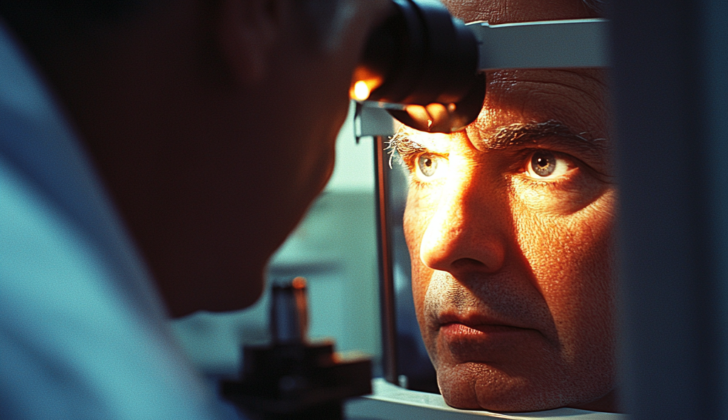
Testing for Pterygium
When examining your eyes, the doctor will perform a comprehensive eye examination. This includes checking your vision (visual acuity), the movement of your eyes (extraocular movements), and looking at the front parts of your eye. They will also perform a detailed test called refraction to determine your precise level of vision impairment or astigmatism, which is an irregular curve of the eye’s lens or cornea.
The doctor will rule out “Dry Eye Syndrome,” a condition when your eyes don’t produce enough tears to keep them comfortably lubricated. This can be determined by the Schirmer’s test that measures tear production or a tear film break-up time test that examines how long it takes for your tears to dry up.
If they suspect that you have a pterygium, a growth on the cornea that can affect vision, they will examine it further. They consider its location, size, the number of blood vessels (vascularity), extent, and the cornea’s area involved. Measurements of the pterygium are taken with a tool called Castroviejo’s calipers. They may use a handheld light (torchlight) that shines obliquely to inspect it better and confirm their investigation with a slit-lamp examination, which magnifies the front part of your eye.
A pterygium has three parts: the cap, which includes a clear area surrounding its leading edge; the head, which is next to the cap; and the body, which is the main part connected to the conjunctiva, a clear tissue covering the front of your eye.
Depending on its characteristics, your doctor may classify the pterygium into two types. Progressive pterygium is thick, flesh-like, vascular and slowly moving towards the center of the cornea, which can affect vision. On the other hand, Atrophic pterygium is thin, lacking fullness, has fewer blood vessels, and doesn’t change or progress much.
Treatment Options for Pterygium
What else can Pterygium be?
When examining someone who may have pterygium, a type of growth in the eye, doctors need to consider other conditions that could cause similar symptoms. These include:
- Pseudo-pterygium: This is an inflammatory reaction where the cornea and the bulbar conjunctiva, or the transparent tissue covering the front of the eye, stick together. The condition is different from pterygium because a probe can pass beneath the tissue. It can show up anywhere in the eye and doesn’t progress or worsen.
- Pinguecula: This is a yellowish growth on the bulbar conjunctiva near the limbus, the border between the cornea and the sclera, the white part of the eye. It may be due to fat, protein, or calcium deposit and often locates on the nasal side. Patients usually don’t experience symptoms but may have mild redness and a sensation of something in the eye when it’s inflamed.
- Nodular Episcleritis: In this condition, a discrete area of episcleral tissue, which lies right under the conjunctiva, becomes inflamed and raised. It is generally mildly painful, a differentiating factor from pterygium. When the eye drop with 2.5% phenylephrine is used, the superficial blood vessels in the eye will blanch or lose their redness.
- Marginale Keratitis: An inflammatory disease that affects the peripheral part of the cornea, the clear, front surface of the eye. It is characterized by an inflamed corneal surface with stromal infiltrates, deterioration of the epithelium, and ulceration that is separated from the eye border by a clear zone.
- Phlycten: This condition results from a delayed immune reaction causing nodular inflammation of the conjunctiva.
- Conjunctival Carcinoma in situ/ Bowen epithelioma: This is a rare condition that presents as a gelatinous mass.
- Ocular Surface Squamous Neoplasia (OSSN): Another type of growth in the eye, which is also looked at in the diagnosis.
Possible Complications When Diagnosed with Pterygium
There are complications that can arise during and after pterygium surgery. Pterygium surgery is a procedure to remove a non-cancerous growth from the white part of the eye.
Complications that occur during the surgery can include excessive bleeding, damage to the muscle on the inside of the eye, wide removal of the pterygium, grafting issues (like improper positioning, size, or thickness of the graft), and accidental puncture of the eyeball with the surgical needle.
After the surgery, the pterygium can sometimes grow back, which is a major complication. Other complications after the surgery can include swelling of the graft, development of a hollow at the surgery site, unhealed surgical spots, overgrowth of granulation tissue, fusing of the eye membranes, graft death, graft shrinkage, and the formation of small sacs of liquid or blood beneath the conjunctiva (the clear, thin membrane that covers the front surface of the eye). Other complications include thinning of the white part of the eye, granulation tissue around the stitches, and much more.
Finally, using medications called MMC and beta radiations, to prevent growth recurrence, has been linked to scleral necrosis (death of the white part of the eye), eye infection, severe secondary glaucoma (a severe increase in eye pressure), and iritis (inflammation of the iris).
Common Complications:
- Excessive bleeding
- Damage to the medial rectus muscle
- Grafting issues
- Accidental puncture of the eyeball
- Recurrence of pterygium
- Swelling of the graft
- Unhealed surgical spots
- Fusing of the eye membranes
- Graft death
- Formation of small sacs of fluid or blood
- Corneoscleral thinning
- Granulation tissue around the stitches
Complications from MMC and Beta Radiations:
- Scleral necrosis
- Eye infection
- Severe secondary glaucoma
- Iritis
Preventing Pterygium
If you have pterygium, a condition where tissue grows over your eye, it’s important to know that it can reappear even after surgery. After the operation, you might be prescribed special eye drops — mitomycin or steroids — that can help reduce this chance. However, it’s crucial to only use these under the guidance of your eye surgeon. If these drops are not used correctly, they could lead to serious eye problems, like the thinning or ‘melting’ of the clear front surface of the eye (cornea) or the white outer layer of the eye (sclera), which can cause vision loss. Incorrect use can also lead to legal issues.
It’s also worth noting that for older patients, a different eye disease called OSSN can look a lot like pterygium. To make sure it’s not OSSN, the tissue removed during surgery might need to go through further checks under a microscope (histopathological examination). This helps ensure that you’re getting the most appropriate treatment for your condition.


