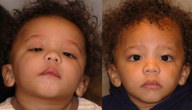
What is Ptosis?
Ptosis is a condition where the upper eyelid droops unusually low, also known as blepharoptosis. This drooping can partially or entirely block vision and can either be present from birth or develop later in life. The correct treatment, whether it’s in the form of medicine or surgery, is dependent on identifying what specifically caused Ptosis. This ensures patient recovery progresses positively.
What Causes Ptosis?
Ptosis, or drooping of the upper eyelid, is typically divided into two main categories – congenital (you’re born with it) or acquired (you get it as you age). The acquired type is usually broken down into five subtypes:
1. Neurogenic: This is when your levator muscle, which lifts your eyelid, doesn’t work correctly because of nerve issues. Various conditions like third nerve palsy, Horner syndrome, Marcus Gunn jaw-winking syndrome, multiple sclerosis, and more could cause these nerve issues.
2. Myogenic: In this subtype, there is problem with either the levator muscle itself or the connection between the nerve and the muscle, called the neuromuscular junction. This can include conditions like myasthenia gravis (a muscle weakness disorder), ocular myopathy (eye muscle disease), simple congenital ptosis, or blepharophimosis syndrome (a condition which causes narrowing of the eyelid opening).
3. Mechanical: Here, the function of the levator muscle is impacted by an outside abnormal structure like a tumor, a chalazion (a lump on the eyelid), a contact lens that’s misplaced, or scarring.
4. Aponeurotic: Also known as Involutional ptosis, it occurs when there’s a problem with the levator muscle’s tendon due to aging, injury, or complications from surgery.
5. Traumatic: This subtype is caused by any kind of eyelid injury that leads to a cut or damage to the levator muscle or the eyelid, or a fracture with reduced blood flow to the top of the eye socket.
In all cases, the result is a drooping of the eyelid, making it difficult for the eye to remain fully open.
Risk Factors and Frequency for Ptosis
Congenital ptosis, a condition where the upper eyelid drops down, is the most frequent kind of ptosis, and it usually occurs more in males. “Simple congenital ptosis” is the specific type that is most common. In circumstances where ptosis is not present from birth but develops later, the most common form is “aponeurotic ptosis,” which typically appears in later adulthood. As of now, there isn’t sufficient data about how often ptosis occurs. However, it doesn’t seem that factors like race impact how prevalent this condition is.
Signs and Symptoms of Ptosis
Ptosis is a condition where the eyelid droops, leading to visual disturbances that can range from mild to severe. These disturbances may be present in one or both eyes and can also cause cosmetic issues. Ptosis may occur with other problems, depending on the cause.
When taking a patient history for ptosis, it’s important to know when the symptoms began and how long they’ve lasted. This can help distinguish between inborn ptosis and ptosis that develops later in life. Other symptoms may be present if the ptosis is linked to another condition, such as differing symptoms throughout the day, double vision, changes in eye position, or fatigue. Information on previous injuries, surgeries, or medical treatments is also useful.
A clinical evaluation for ptosis involves checking for a drooping lid that covers more than 2mm of the cornea, which causes the eyelid opening to be narrower than usual. Other indicators include a backwards tilt of the head, forehead wrinkles, or raised eyebrows, which are signs the patient is trying to compensate for the drooping lid. The presence of scarring, swelling, or other abnormal structures near the eyelids should also be checked. Eyeball changes, like changes in position or bulging, may also be present.
A series of clinical signs can aid in diagnosing ptosis:
- A missing upper lid crease may indicate that the ptosis is congenital, or present from birth.
- Pupil function should be checked for signs of Horner syndrome, characterized by small pupils, and cranial nerve III palsy, which results in larger pupils.
- Eyeball movement should be evaluated for signs of nerve damage.
- The jaw-winking sign, a jerking movement of the upper lid when the jaw moves, needs to be examined to exclude Marcus Gunn jaw-winking syndrome.
Before ptosis surgery, the flow of tears and the reflexes of the eye need to be checked. A test called the phenylephrine test may be used to evaluate the muscle responsible for lifting the eyelid. In this test, phenylephrine is placed in the eye, and a positive response is seen if the eyelid lifts. This indicates that the patient is a good candidate for a specific type of surgery.
In some cases, ptosis can be a symptom of a disorder called myasthenia gravis, which can either impact the entire body or just the eyes, resulting in drooping eyelids that spare the pupils. This can be tested using an edrophonium test, in which the drug edrophonium is administered, and a positive result is the lifting of the eyelid within 2 to 5 minutes. Because this test isn’t particularly sensitive, another test involves placing an ice pack over the drooped lid for 2 to 5 minutes and checking for improvement.
Testing for Ptosis
For a condition known as blepharoptosis, which is a fancy word for drooping of the eyelid, doctors will look at several things to figure out the best treatment:
1. Levator muscle function: This is a muscle that helps lift your eyelid. To see how well yours is working, a doctor will put their thumb on your eyebrow while you look down, then measure how far your eyelid moves when you look up. Normal movement is about 15 millimeters (mm), and anything less than that may suggest that your levator muscle is not working properly. This information is important as it helps determine the best surgical technique for treating the droopy eyelid.
2. Margin-reflex distance: For this, you look at a light, and the doctor measures the distance between the reflection of that light on your eye and the edge of your upper eyelid. Typically, this distance should be between 4 to 5 mm.
They’ll also measure the distance between your upper and lower eyelids. Normally, your upper eyelid covers about 2mm and your lower eyelid covers about 1mm of your eye’s cornea. The normal distance between these two eyelids is 7 to 10 mm in men and 8 to 12 mm in women. This can help them figure out if you have a mild (1 to 2 mm), moderate (3 to 4 mm), or severe (4 mm or more) case of blepharoptosis.
Additionally, your doctor may measure the distance from the edge of your eyelid to the crease of your eyelid when you’re looking down. This measurement in men is usually 7 to 8 mm, and in women, it’s 8 to 10 mm. A higher than normal measurement could suggest a particular type of ptosis, while an absent or vague measurement could imply that the ptosis is congenital (something you were born with).
If your doctor suspects a condition called myasthenia gravis, they may order blood tests for specific antibodies that cause this autoimmune disorder. However, keep in mind that these tests are only positive in about 50% of people with eye-related myasthenia.
Sometimes, doctors will order thyroid tests. Although rarely droopy eyelids may be a sign of an underactive thyroid, and it’s also been noted that there might be a link between thyroid disorders and myasthenia.
Lastly, your doctor might recommend an X-ray or a CT/MRI scan of your brain or eye area if they think you might have a tumor or a nerve defect, trauma, or other conditions.
Treatment Options for Ptosis
Treatment for a drooping eyelid, or ptosis, depends on what caused it, how much the eyelid is sagging, and how well the eye’s levator muscle, or “lid lifter,” is working. In some cases, such as when a drooping eyelid is caused by a lump called a chalazion, simply removing the lump can fix the problem. However, usually, the main treatment involves surgery. In more specific cases, there are some non-surgical options available.
Surgery is typically necessary for ptosis that’s present from birth (congenital ptosis) or other types of ptosis that don’t get better with non-surgical treatment. What the surgery involves depends on what’s causing the ptosis and what the eye doctor determines before the procedure.
One surgical method is levator resection, which shortens the levator muscle if it’s not fully paralyzed and the ptosis is mild (2 mm) to moderate (3 to 4 mm). Different techniques are used to do this, like the Everbursch method (through the skin), the Blaskovics method (through the palpebral conjunctiva, which is the tissue that covers the inside of the eyelids), and the Fasanella-Servat method (which removes a part of the tarsal plate – a plate of dense connective tissue – along with the palpebral conjunctiva and the levator muscle and is usually used for mild ptosis).
There are also procedures such as the Motais procedure (using the superior rectus muscle to lift the eyelid if the levator muscle is paralyzed) and the Hess’s procedure (using the forehead muscle to raise the eyelid if both the superior rectus and the levator muscles are paralyzed)
In cases of severe ptosis (over 4 mm) with poor levator muscle function, especially in children who have had ptosis since birth, a frontalis brow suspension may be done. Here, the eyelid is linked to the forehead muscle with a structure from the fascia lata (a dense fibrous structure in the thigh) or a synthetic material like mersiline mesh to help lift the eyelid.
Another method, known as aponeurotic strengthening, advances the aponeurosis- the white section of the muscle, mainly used in age-related ptosis.
In certain cases, such as in myogenic (muscle-related) and some types of neurogenic (nerve-related) ptosis, non-surgical treatment might be useful. For example, taping the upper eyelid so that it stays open or adding small support devices, known as eyelid crutches, to glasses.
What else can Ptosis be?
Pseudoptosis is a condition that may be mistaken for ptosis, a drooping or falling of the upper eyelid. Several situations can cause this mistake:
- A retracted lid on the opposite eye
- The upward movement of the same eye followed by the eyelid
- Skin hanging over the upper lid
- The drooping of the brow itself
- Swelling of the upper lid, which can be caused by conditions such as preseptal cellulitis, orbital cellulitis, or a chalazion (a lump in the eyelid)
- A smaller than usual eye size or eye socket, as seen in conditions like microphthalmos, phthisis bulbi, enophthalmos, etc.
What to expect with Ptosis
The outcome of ptosis, a condition where the upper eyelid droops, largely depends on the type of ptosis and treatment received. If the right surgical procedure is used based on a thorough examination before surgery, patients usually see great improvements. For instance, if a ptosis patient has weak levator muscles (the muscles that lift your eyelid) and a levator resection procedure (where part of this muscle is trimmed and stitched back to tighten the eyelid) is performed instead of a fascia lata sling procedure, the outcomes might not be as good.
A well-conducted preoperative assessment is necessary to decide on the right procedure. Close monitoring and proper care after the surgery can also greatly enhance the success of the surgery and improve the condition of drooping eyelids, regardless of the type of ptosis.
Possible Complications When Diagnosed with Ptosis
After an operation to correct droopy eyelids – called ptosis repair – there can be both early and late complications.
The most common early complications include:
- Inconsistencies in the height and shape of the eyelids
- The eyelids being corrected too little (under-correction) or too much (over-correction), which usually improves over time
- Less common, these complications can also include infections at the site of surgery, bleeding, eyelid not fully closing (lagophthalmos), and exposure keratopathy – a condition affecting the outer surface of the eye that needs careful monitoring
Generally, incomplete closure of the eyelid, or lagophthalmos, resolves by itself within 2 to 3 weeks.
On the other hand, late complications of ptosis repair can last longer and include:
- Continued trouble with eyelid closure
- Exposure keratopathy that can get worse
- Reoccurence of the droopy eyelid condition – ptosis
- Persistent overcorrection or undercorrection, which might need another surgery to address
Preventing Ptosis
It’s vital for patients to understand what causes ptosis, a condition where the upper eyelid droops, and what might happen if it’s left untreated. There are various treatment options available, both surgical and non-surgical, and it’s important to discuss all these options to decide the best one based on an individual’s needs and health condition. The doctor should also explain to the patient about potential complications that might occur after surgery, either immediately or in the future, and how these would be managed.


