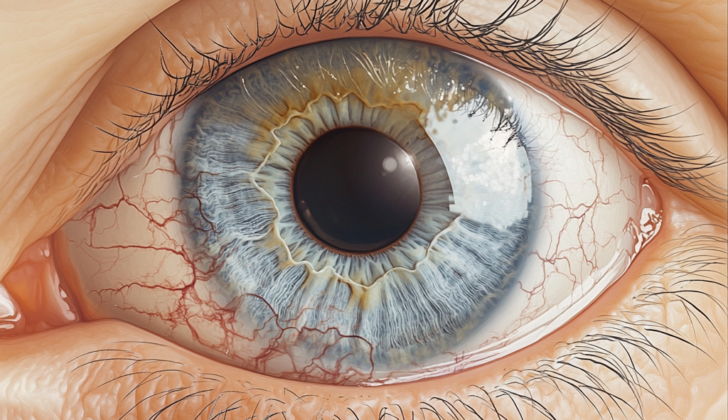
What is Salzmanns Nodular Corneal Degeneration?
In 1925, an Austrian eye doctor named Maximilian Salzmann first described a condition where lumps grow on the eye following certain types of inflammation in the cornea, the clear layer at the front of the eye. Over time, Salzmann’s Nodular Degeneration (“SND”) was further clarified as a condition where fibrous, non-inflamed lumps grow in any area of the cornea, but often around the edges. Although we don’t always know why, these lumps can form due to inflammation, injury, or surgery.
SND is uncommon and usually happens when patients are in their 50s. However, patients as young as 4 and as old as 90 can have this condition. Most times, SND occurs in both eyes and it is seen more often in women and among Caucasians. Unfortunately, the lumps don’t typically disappear spontaneously, but the symptoms can be eased with treatments like eye lubricants and topical anti-inflammatory medications.
In more resistant cases where the symptoms aren’t improved by these treatments, some surgical options can be considered: surgically removing the lumps, performing a procedure to reshape the cornea (“phototherapeutic keratectomy”) with or without a specific medication (“mitomycin-C”), or replacing part or all of the cornea (“lamellar or penetrating keratoplasty”). Even though patients may not feel any symptoms, SND can negatively affect eye health and vision.
The outlook is generally positive for patients with SND, but identifying and treating the condition and any underlying factors are vital. There have been no reported cases of SND clearing up spontaneously without treatment.
What Causes Salzmanns Nodular Corneal Degeneration?
Certain conditions affecting the eyes, such as dry eye, chronic blepharitis, a type of inflammation of the eyelids, long-term wearing of contact lenses, and injuries, can increase the risk of developing Salzmann nodular degeneration (SND), which is a type of eye disease characterized by nodules, or small lumps, forming on the eye.
One study showed that wearing contact lenses accounted for about a third of the cases of SND. It is thought that this may be due to the way contact lenses can interfere with the normal renewal of cells in the eye, slow down the rate at which the cells in the center of the cornea, or the clear front part of the eye, are shed, and destabilize the tear film that normally protects the cornea.
Eye surgeries that involve cuts or wounds to the cornea, including cataract surgery, radial keratotomy, a type of eye surgery for nearsightedness, penetrating keratoplasty, which is a corneal transplant, or LASIK, a type of refractive eye surgery, can also increase the risk of developing SND. Of 180 eyes with SND evaluated in a 2010 study, more than a quarter had a history of precursor eye surgery. Post-LASIK SND may happen due to dry eye disease that develops after the surgery, irregular corneal tissue in the area of where the LASIK flap was created, or simply the trauma caused by the surgery itself. While these post-LASIK nodules often respond to simple treatments, there have been reported cases where a superficial keratectomy, a type of eye surgery, was needed.
Although there has not been a specific gene found to be associated with SND, there have been cases reported where the condition has appeared in several generations of a family, suggesting that there could be a heritable aspect to the disease. It’s possible that a unique genetic factor or an defect, or mutation, in the TGF-β gene, which is known to play a role in other corneal diseases, may be involved.
Risk Factors and Frequency for Salzmanns Nodular Corneal Degeneration
Salzmann nodular degeneration (SND) is an uncommon condition. This typically occurs in people between the ages of 50 to 60, with the average age being just a little over 60. The condition has been observed in individuals as young as 13 and as old as 92. Interestingly, it has been found to appear more frequently in certain age groups, particularly people in their 50s or 80s.
It has been noticed that SND affects women more often. In fact, women made up 78% of the cases in the original study of the condition, and this trend has been replicated in later research.
Sometimes, SND can affect both eyes, happening in just over half of all instances. But it’s important to note that it could be more severe in one eye than the other.
Interestingly, SND has also been found in patients with Crohn’s disease. While there are only a couple of confirmed cases, there have been several mentions of eye nodules in individuals with this condition.
- Salzmann nodular degeneration (SND) often affects people aged 50 to 60.
- The average age of patients with this condition is around 60.8, but it has been seen in people aged 13 to 92.
- Women are more likely to have SND, accounting for 78% of cases in several studies.
- Over half of the time, SND affects both eyes, though the severity might differ between eyes.
- SND has been noted in some cases of patients with Crohn’s disease.
Signs and Symptoms of Salzmanns Nodular Corneal Degeneration
Salzmann nodular degeneration (SND) is an eye condition where only a minor portion of patients experience symptoms. When symptoms do occur, they can include feelings of having a foreign body in the eye, irritation, excessive tearing, or sensitivity to light. In some studies, a decrease in visual acuity, or the sharpness of vision, was the most common symptom reported. However, patients usually only experience a slight reduction in vision.
In other research, visual disturbances were the prevalent reason people opted for surgery, accounting for 85% of cases. The nodules’ location can impact the primary complaint. If the nodules are located centrally, patients often report decreased or distorted vision. On the other hand, nodules located more towards the edges of the eyes are usually linked with the sensation of a foreign body in the eye.
Visual distortions can occur due to various causes such as the involvement of the nodules with the visual axis, uneven astigmatism, far-sightedness, or issues with the tear film. Peripheral nodules may also cause the central part of the cornea to flatten. This could lead to increased far-sightedness, and every quarter of the eye that’s affected can increase astigmatism by 0.38 D.
Testing for Salzmanns Nodular Corneal Degeneration
Salzmann nodular degeneration (SND) is a rare eye condition that causes small nodules or bumps to develop on the surface of the cornea, which is the clear, dome-shaped tissue on the front of your eye. The condition normally occurs as a result of an injury, surgery, or long-term eye inflammation. It’s diagnosed primarily with a slit lamp exam where a doctor uses a special microscope to examine your eyes.
Doctors may also use other tools such as high-frequency ultrasound biomicroscopy (UBM), in-vivo confocal microscopy (IVCM), optical coherence tomography of the anterior segment (AS-OCT), or corneal topography and tomography to support their diagnosis. These are advanced imaging techniques that allow doctors to take detailed pictures of different parts of your eye.
When examining your eyes, the doctor may see nodules that are normally smaller than 2mm but can be as large as 4×2.5 mm. The nodules can appear anywhere on the cornea, with the location depending on the cause of the condition. For example, nodules are often found in the middle of the cornea in people who have had LASIK eye surgery, or at the 3 and 9 o’clock positions in people who have a history of wearing contact lenses. The nodules are usually greyish-blue, round, and are located towards the outer part of the cornea rather than the center.
Interestingly, one study showed that each nodule corresponded to an area of new blood vessel growth or neovascularization in the cornea, known as a stromal pannus, that did not enter the nodule. In another study, an interesting pattern called the Bell phenomenon was found to occur in patients depending on the location of their nodules.
In addition, these eye scans can provide detailed images of your cornea. For example, using UBM, the doctor can see that the nodules are made up of a thin layer of epithelial cells overlying a reflective substance. It’s also notable that corneal topography, a tool for mapping the surface of your cornea, can yield incorrect results due to the presence of the nodules.
The AS-OCT is another non-invasive imaging technique that is useful for the diagnosis and management of SND. AS-OCT can provide ‘optical biopsies,’ which are essentially virtual snapshots of the cornea that can be used to understand the disease without needing to take any tissue samples. These images typically show bright, reflective deposits underneath the epithelial layer of your cornea.
Likewise, IVCM allows doctors to analyze the tiny structures within the nodules. Under IVCM, regular corneal cells appear arranged like honeycomb shapes, but the cells within the nodules appear irregular, including elongated, polygonal cells of varying sizes. Deeper within the nodules, the keratocytes – cells within the stroma or ‘body’ of your cornea, appear to be more active whereas the overall corneal structure within the nodule appears unstructured, with an increase in reflective background. However, the Descemet membrane and the endothelium, which are deeper layers of the cornea, appear normal.
Treatment Options for Salzmanns Nodular Corneal Degeneration
The treatment method for nodules, which are small, abnormal lumps, depends on their size, location, and how severe the symptoms are. If a patient doesn’t have symptoms or the symptoms are mild, doctors usually opt for a “wait-and-see” approach or simple care methods. However, severe cases may need surgery, especially if it’s affecting a patient’s vision. Sometimes, if the nodules don’t improve with simple care measures, surgery might be necessary. Patients who have nodules in many areas may also need surgery.
Simple Care Measures
Many people find relief with simple care strategies. This can include using lubricants, warm compresses, and gently massaging the eyelid. Keeping the eyelids clean can help improve dry eye symptoms. Most people can avoid surgery with these strategies. Initial treatment usually includes drops to moisten the eyes. Other medicines that might be used include eye drops that contain steroids, nonsteroidal anti-inflammatory drugs, immunomodulators (drugs that can modify the immune response), and a drug named doxycycline. After a certain type of eye surgery (LASIK), plugs for tear ducts have been used along with certain medications, with symptoms improving after six months. If nodules are causing problems in those who wear contact lenses, stopping or reducing lens wear may help. The fit of contact lenses should be checked.
Nodulectomy/Superficial Keratectomy
The most straightforward surgical approach is to cut out the nodule and remove the thin layer of the cornea (the clear front part of the eye). The surgeon removes the neighboring epithelium (the outer layer of cells) or debridement (removal) can be carried out. The surgeon lifts the edge of the nodule and creates a surgical plane, which is a gap that makes the removal easier. If the surgeon cannot create a surgical plane, special tools may be used to create one.
If the nodule is stuck to the limbus (the border between the clear front part of the eye and the white part), scissors might be needed. After the nodule is removed, the underlying stroma (the thicker, middle layer of the cornea) is usually regular and smooth. However, in more complicated cases, other problems may make surgery more challenging.
Some patients have irregularities left in their Bowman layer (the second layer of the cornea). These irregularities can be treated with a specific tool or a procedure called Phototherapeutic Keratectomy (PTK). There is a higher probability of recurrence in cases where these defects are deeper. Some cases have been treated with superficial keratectomy followed by AMT (applying a special type of tissue), but recurrences were reported. Data on such specific cases is limited.
A drug named Mitomycin-C (MMC), which can stop DNA synthesis and induce cell apoptosis (programmed cell death), has been found to be helpful. When applied during surgery, MMC can stop the progression of Salzmann nodules and prevent recurrence after removal. One study showed no recurrence for an average of 28±15 months following this technique.
Keratoplasty
Rarely, a surgical procedure called lamellar keratoplasty (LK), a type of corneal transplant, may be needed to treat Salzmann’s Nodular Degeneration (SND). The change in visual acuity (sharpness of vision) was found to be similar between groups treated with automated lamellar keratoplasty (ALK) and Phototherapeutic Keratectomy (PTK), but the PTK group had fewer complications. Moreover, some cases of SND have been reported to recur after keratoplasty. Full thickness cornea transplant (PK) is rarely needed, usually only in cases with extensive additional diseases or if the cornea is accidentally punctured during LK.
Additional Considerations
For the most accurate results for cataract extraction, SND should be removed beforehand. If not, it may affect the measurements taken before cataract surgery and consequently affect the power calculations of the Intraocular Lens (IOL), the artificial lens that replaces the eye’s natural lens during cataract surgery.
What else can Salzmanns Nodular Corneal Degeneration be?
Salzmann nodular degeneration, or SND, is a condition that affects the cornea – the clear front surface of the eye. However, it can be quite similar to other conditions, so doctors have to carefully evaluate a patient’s symptoms to ensure they make the correct diagnosis.
Here are some conditions that can look like SND, along with some of their unique features that can help doctors tell them apart:
- Bullous Keratopathy: Usually shows signs of membrane folds, swelling in the cornea, or increased eye pressure. Unlike SND, these nodules contain water.
- Climatic Droplet Keratopathy: This condition often presents with golden-yellow or clear droplets in the area of the cornea that’s exposed when we blink.
- Corneal Amyloidosis: This involves the buildup of protein deposits called amyloid in the central region of the cornea. These deposits are usually under the surface layer of the cornea.
- Primary Amyloidosis: Usually shows a mound of amyloid in the cornea, and these deposits are typically subepithelial.
- Secondary Amyloidosis: This tends to appear in areas that have been injured before, presenting as yellow-white to pink waxy nodules.
- Corneal Keloid: These changes are typically seen in younger age groups and can be due to inflammation, injury, or associated with specific health conditions such as Lowe syndrome.
- Hereditary Hypertrophic Scarring: Involve nodular scaring after minor traumas or surgeries and can be difficult to differentiate from SND.
- Peripheral Hypertrophic Subepithelial Corneal Degeneration: This condition causes corneal opacities to appear in the areas of the cornea that are exposed during blinking. These changes are often seen in both eyes and involve both the near (nasal) and far (temporal) sides of the cornea.
What to expect with Salzmanns Nodular Corneal Degeneration
The outlook is generally positive with treatment focused on addressing the root cause. Even though some people who show no symptoms can be carefully watched without seeing the condition get worse, there haven’t been instances where the condition disppeared on its own. Different studies show different rates of the condition coming back.
Nonetheless, treating the root cause of the condition can lower the chances of more nodules forming in the future.
Possible Complications When Diagnosed with Salzmanns Nodular Corneal Degeneration
Salzmann nodular degeneration, also known as SND, can lead to severe complications. Some of these complications include:
- It can cause irritation to the surface of the eye, resulting in sensitivity to light (photophobia)
- Excessive tearing
- Involuntary closure of the eyelid (blepharospasm)
- Repeated damage to the clear tissue on the front of your eye (corneal erosions)
- The nodules of Salzmann can flatten the eye’s clear surface (cornea), causing irregular eye shape (astigmatism). In some cases, if the nodules are removed, the astigmatism can be fixed.
Preventing Salzmanns Nodular Corneal Degeneration
Salzmann nodular degeneration (SND) is a condition that affects the eye. It often develops due to conditions such as meibomian gland disease (MGD), which affects the oil glands in the eyelids, dry eye, and extended use of contact lenses. These factors are the most commonly mentioned in the development of SND.
If you are showing early signs of eye inflammation, which could lead to SND, it’s crucial to take preventive measures. These include regularly applying preservative-free lubricant eye drops to keep your eyes moist, maintaining clean eyelids to prevent the accumulation of debris that may irritate the eyes, and avoiding overuse of contact lenses. By following these steps, you could potentially reduce the progression of this condition.


