What is Blount Disease (Bloom-Torre-Machacek Syndrome or Congenital Telangiectatic Erythema)?
Blount disease, also known as ‘bowed-leg’ or tibia vara, is a condition seen in children where the shinbone or tibia grows unevenly which makes the leg bend outwards like a bow. This condition is caused by uneven growth of the cartilage in the upper part of the shinbone, located on the inner side. What happens here is that excessive pressure forces result in irregular bone formation. Blount disease can affect one or both legs and it could show up in two ways based on the child’s age – infantile (early-onset) or adolescent (late-onset).
The early-onset form of Blount’s disease is more common, usually appearing in children aged between 1 and 5, often affecting both legs and becoming more pronounced when the child starts to walk. The late-onset form shows up in older children and it could affect one or both legs.
While obesity (being overweight), starting to walk early, and being of African-American descent are all factors that can increase the risk of developing Blount disease, we do not yet fully understand the exact cause of the condition. The severity ranges from irregularities in the surface of the joint to differences in leg length. Treatment for Blount disease depends on the child’s age and the severity of the condition at the time it is found. It could range from putting a brace on the leg to slow the growth and straighten the bow to surgical procedures.
X-ray findings are the main way to diagnose Blount disease. There is a classification system known as the Langenskiöld classification system that outlines the 6 stages of Blount disease based on X-ray images. This condition was first recognized by Walter Putnam Blount, an orthopedic surgeon working with children, in 1937.
What Causes Blount Disease (Bloom-Torre-Machacek Syndrome or Congenital Telangiectatic Erythema)?
Blount disease, a condition that affects the growth of the shin bone, is caused by a combination of biological and physical factors. Although excessive physical strain on the shin bone is a major factor, especially in children who are overweight and start walking early, it’s not the only cause.
The infantile form of the disease, which also occurs in children of normal weight, and a higher rate of the disease in African-American children, suggest that there may also be a genetic link. So, apart from physical strain, genetic factors also play a role in the development of Blount disease.
Risk Factors and Frequency for Blount Disease (Bloom-Torre-Machacek Syndrome or Congenital Telangiectatic Erythema)
Blount disease is quite rare in the United States, affecting less than 1% of the population. The infantile form of this disease is more common and affects males more often than females. Most notably, in 80% of these cases, it affects both sides of the body. The adolescent form of Blount disease, also known as adolescent genu varum, is usually less severe and typically only affects one side of the body. This condition is most commonly found in children of African and Scandinavian descent.
Signs and Symptoms of Blount Disease (Bloom-Torre-Machacek Syndrome or Congenital Telangiectatic Erythema)
Bowed legs, or genu varum, is a common condition seen in children until they’re about two years old. After this point, their legs gradually shift into a position called valgus until they’re around three years old. If a child is bigger and has bowed legs that continue past two years old, it might be the first sign of a growth disorder. As the disorder develops, the shape and alignment of the knee may change, not just causing their legs to bow but also possibly their tibia to twist and for one leg to be longer than the other.
Blount’s disease is a particular type of growth disorder, and it can show up at different times in a child’s life. The version that appears early, Infantile Blount disease, is typically spotted in children between the ages of one and three. This condition normally affects both legs and causes the tibia (shin bone) to twist inwards, making the legs bow. Pain is usually not a symptom, but a noticeable bump might appear at the inside edge of the knee. If the child’s knee shifts to the side when they put weight on it, it indicates a more serious issue that has developed by around ages six to eight. From this point, less invasive treatments aren’t likely to work.
A later version of Blount’s disease, called Adolescent Blount disease, may appear in late childhood or early adolescence. It often causes pain on the inside of the knee and usually only affects one leg. This form of Blount’s disease particularly affects kids who are bigger or overweight and sometimes can cause irregularity in the thigh bone close to the knee.
Testing for Blount Disease (Bloom-Torre-Machacek Syndrome or Congenital Telangiectatic Erythema)
Blount disease is a condition that affects the growth of the shin bone and can often be diagnosed through a detailed health history, physical examination, and X-rays. For the early stages of the disease, doctors typically use X-ray images of the entire leg in a standing position to look for signs and measure the degree of inward bending (varus). It is essential that these images capture the area from the hip down to the ankle on both sides.
There are specific markers doctors look for that suggest Blount disease. These include deformities on the inner side of the growth plate of the shin bone, an enlarged and uneven growth plate, unusual hardening of the bone tissue, and a sloping of the ends of the bone towards the inner side.
Doctors use several different measurements of angles in the knee joint to diagnose Blount disease in children. The Levine-Drennan angle, which measures the relationship between the bone shaft and its upper growth plate, is one such measurement. If the measured angle exceeds 11°, it often indicates the presence of Blount disease.
Another important angle is the metaphyseal-diaphyseal angle (MDA). The MDA predicts the progression of Blount disease and involves assessing the intersection between a line drawn from the sharpest point on the inner and outer sides of the tibial metaphysis (the growing end of the bone) to a line at a right angle to the length of the tibial diaphysis (the shaft of the bone). Angle measurements can suggest the likelihood of the disease becoming worse over time or the likelihood of spontaneous recovery.
Magnetic resonance imaging (MRI) can be very useful in this diagnosis as it is particularly effective in assessing the cartilage, menisci, ligaments, and blood supply to the growth plate. MRI is also better than X-rays in detecting changes in the cartilage. Certain types of MRI scans can be especially beneficial for children who have a delayed or overlooked form of Blount disease that isn’t seen until after age 4 but before the growth plate has turned into bone.
The severity of Blount disease is divided into six stages using the Langenskiöld Classification System, which is specifically used for the infantile form of the disease. These stages represent increasing levels of severity and collapse of the inner growth plate, with an unusual bone formation becoming evident from stage five onwards. This unusual bone formation, also known as a physeal bar, can cause angulated deformities and differences in limb length in children who are still growing. Although MRI-based classification systems have recently gained popularity, X-ray-based classification remains the most widely used method.
Finally, there are four recognized stages of ligament laxity, or looseness, that doctors look out for:
- Stage 0: Normal laxity
- Stage +: Inner side looseness
- Stage ++: Outer side looseness
- Stage +++: Looseness in multiple directions
Treatment Options for Blount Disease (Bloom-Torre-Machacek Syndrome or Congenital Telangiectatic Erythema)
The treatment for Blount’s disease, a growth disorder that causes the lower leg to turn inward, resembling a bowling pin, can vary and depends on the age of the child and the severity of the leg bend. Treatment is necessary to prevent further damage to the leg joint and to ensure both legs are of equal length when the child reaches full growth.
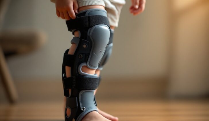
For children diagnosed before age 4, wearing a knee-ankle-foot orthosis (KAFO) brace might be recommended. This type of brace runs from the upper thigh down to the foot, applying a force that helps straighten the knee. This treatment is usually more successful when it begins before the child is 3, particularly in thinner kids, and is mostly worn at night. If the brace doesn’t work, the doctor might recommend a surgical procedure called an osteotomy.
For children who are still growing, there’s another surgical option called hemiepiphysiodesis, or guided growth. This procedure slows down the growth on one side of the leg, allowing the other side to catch up and gradually straighten the leg. The procedure involves inserting plates, pins, or staples into the leg bone near the growth plate. One advantage of this procedure is that once the hardware is removed, the leg can continue to grow normally. However, this treatment requires at least 4 years of remaining growth to be effective.
An osteotomy procedure could be recommended for children under 4 with progressive Blount’s disease. This procedure involves cutting and reshaping the bone to correct its alignment. The surgery aims to achieve a slight outward bend to counteract the inward curve of the disease. Various osteotomy techniques have been described, and they can result in either an immediate or gradual correction of the leg’s alignment. However, the surgery carries risks, such as nerve damage and severe muscle swelling (compartment syndrome).
If the disease is severe, other types of surgical procedures might be considered. One of these is called a physeal bar resection, which aims to restore normal growth and prevent further deformity. However, this procedure is usually only suitable for children aged under 7. Another type of surgery called medial tibial plateau elevation might be recommended for older children with advanced disease. This procedure involves lifting the inner part of the knee joint to correct its position and prevent a bow-legged gait.
Remember, the treatment strategy for Blount’s disease will be child-specific and will depend upon various factors like the severity of the condition, the child’s age, and their overall health status. The goal will always be to improve the quality of life for the child and prevent future complications. Always consult the treating physician or a medical professional for more personalized advice.
What else can Blount Disease (Bloom-Torre-Machacek Syndrome or Congenital Telangiectatic Erythema) be?
It can be tough to tell the difference between infantile tibia vara (a condition known as Blount’s disease) and normal bowing of the legs in babies. Normally, both the thigh bone (femur) and shin bone (tibia) develop a gentle curve as the child grows. But in Blount’s disease, the curve in the shin bone near the knee is more sudden. If the shin bone angles more than 11 degrees away from the thigh bone, this could be a sign of Blount’s disease.
There are also other conditions to consider that might explain a child’s bowed legs:
- Rickets – a condition that weakens bones
- Ollier disease – a rare bone disease
- Injury to the growth plate in the shin bone near the knee caused by trauma, radiation, or infection
- Osteomyelitis – a bone infection
- Metaphyseal chondrodysplasia – a bone development disorder
- Thrombocytopenia absent radius syndrome – a rare genetic disorder
Blount’s disease often involves asymmetrical beak-shaped bulges and sharp angular deformities in the bone that you don’t see in rickets. Ollier disease differs from these conditions as it results in the formation of multiple noncancerous growths called enchondromas in the bone.
What to expect with Blount Disease (Bloom-Torre-Machacek Syndrome or Congenital Telangiectatic Erythema)
The future outcome (prognosis) of Blount disease significantly relies on the patient’s age and how severe their condition is when they first seek medical help. Blount disease in infants usually has a promising outlook, and there’s a low risk of the bone deformity coming back if treated promptly. It’s even possible for the bone to partially or completely go back to its normal shape if treatment is started early.
However, if treatment is delayed until the more advanced stages of the disease, the condition is likely to get worse. Similarly, patients with a late-onset form of Blount disease might see their condition progress and their joints become significantly deformed if they do not receive treatment.
Possible Complications When Diagnosed with Blount Disease (Bloom-Torre-Machacek Syndrome or Congenital Telangiectatic Erythema)
Complications of certain diseases might cause a deformity to reappear or joint degeneration.
Likewise, surgery might have its own set of complications. These could consist of:
- Deep vein thrombosis, which is a blood clot in deep veins usually occurring in the lower leg or thigh
- Vascular impairment, or issues with blood flow
- Pathologic fractures, which are fractures in weakened bones due to disease
- Wound infection
- Malalignment, which is incorrect positioning of body parts
- Compartment syndrome, a serious condition that involves increased pressure in a muscle compartment
- Premature closure of the growth plate, potentially leading to shorter limbs
- Abnormally fast growth after a period of slow growth
- Migration of surgical hardware, or moving from its initial place


