What is Esophageal Hematoma?
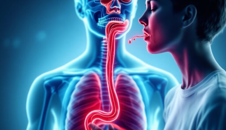
The esophagus is a roughly 10-inch long muscle tube that links your throat to your stomach. If you were to look at it closely, you’d see several layers. The innermost layer is the mucosal, followed by the submucosal that connects the mucosal to the protective muscular layer. The outermost layer is called the adventitial layer. The submucosal layer houses blood vessels, nerve networks known as the Meissner nerve plexus, and esophageal glands.
Sometimes, an unusual and severe health issue can occur called an intramural hematoma of the esophagus, or a dissecting intramural hematoma. In layman’s terms, this means that due to an injury to the mucosal and submucosal layers, blood can collect amongst these layers. This situation can happen on its own or it may result from an injury, like swallowing a foreign object or ingesting harmful substances. It can also happen due to medical procedures.
Patients with an intramural hematoma of the esophagus might have symptoms that look like other serious heart or lung illnesses. Typically, the symptoms include sudden chest pain, discomfort or difficulty swallowing (known as odynophagia or dysphagia), and vomiting blood (known as hematemesis). With a correct diagnosis and proper treatment, patients usually recover excellently from this condition.
What Causes Esophageal Hematoma?
An esophageal hematoma is a blood clot that forms within the wall of the esophagus, the tube that connects your mouth to your stomach. This can occur when there’s a sudden change in the pressure inside your chest and esophagus, which can happen during events like vomiting, severe coughing, or retching. This results in the blood clot, which may then burst into the hollow part of the esophagus. Using medications that thin your blood or stop blood clots from forming, or having conditions that affect your blood’s ability to clot, can all increase the risk of developing this kind of blood clot in the esophagus. In some cases, these esophageal blood clots can form for no clear reason.
This kind of esophageal blood clot can have many causes, and it’s typically categorized as either being spontaneous or as a result of trauma or medical procedures. Trauma-related causes include swallowing foreign objects, doing the Valsalva maneuver (a specific way of breathing out), lifting heavy weights, swallowing a large amount of food in a hurry, having a feeding tube put in your nose, or undergoing certain types of ultrasounds or procedures on your upper gastrointestinal tract like an endoscopy or a specialized procedure called an endoscopic retrograde cholangiopancreatography. Other causes can be due to endotracheal intubation (having a tube put in your throat to help you breathe), and a transesophageal echocardiogram (a type of ultrasound that looks at your heart).
Risk Factors and Frequency for Esophageal Hematoma
Injuries to the esophagus, like trauma and puncture wounds, are quite rare. This makes it hard to pinpoint just how often they occur, especially a case known as an intramural hematoma of the esophagus. A majority of such incidents have been reported in individual case studies. A broad review of many of these case studies shows that patients are most often around 45 or 75 years old. It’s more commonly seen in older women, who are twice as likely as men to develop this condition. The reasons for this are still not clear.
- People with blood disorders, like hemophilia, or who are taking blood-thinning drugs, are more likely to develop an intramural hematoma of the esophagus.
- This type of esophagus problem is relatively rare, but doctors are detecting it earlier now thanks to advancements in modern imaging and inspection tools.
Signs and Symptoms of Esophageal Hematoma
An esophagus’s intramural hematoma can usually be identified when you suddenly experience chest pain or a feeling of pain behind the breastbone. About 80% of patients experience at least two of the three main symptoms, which are:
- Pain behind the breastbone (84% of patients)
- Difficulty swallowing or painful swallowing (59% of patients)
- Vomiting blood (56% of patients)
When doctors go over your medical history, they will ask about any blood disorders, or if you’re taking any blood-thinning medication. They may also check if you have a history of intense vomiting, retching, or any medical procedures involving the esophagus. In rare instances, the condition may be related to swallowing a foreign object.
Physical examinations might not show anything out of the ordinary apart from a rapid heartbeat or pale complexion. It’s crucial, though, to distinguish an esophageal hematoma from other reasons that could cause acute chest pain. If you have trouble swallowing or experience pain when swallowing, this can help rule out severe heart-related causes of pain behind the breastbone.
Testing for Esophageal Hematoma
To diagnose a condition that affects the esophagus (the tube that connects your throat to your stomach), various methods can be used. Usually, the first option is a non-invasive procedure called a computed tomography (CT) scan with intravenous contrast (a dye injected into the veins to make the scanned images clearer). This procedure is typically quick, readily available in most health centers, and will usually show a thicker wall of the esophagus. In instances of larger swellings, it might look as if the tube has disappeared on the scan.
The CT scan, especially when done with contrast, helps doctors understand the position of the esophagus in relation to the surrounding structures like the aorta (the main artery in the body) and other tissues in the middle part of the chest. Sometimes, a liquid is given to the patient to swallow before the scan when there’s a suspicion that there might be a hole in the esophagus wall. If there’s indeed a hole, the liquid will leak out, showing doctors precisely where the damage is.
Other investigations, like endoscopic ultrasound and magnetic resonance imaging (MRI), can also be used to help diagnose an intramural hematoma of the esophagus (a bleeding and swelling within the wall of the esophagus). Endoscopy (where a small camera is inserted down your throat) should wait until doctors are sure there is no hole or tear in the esophagus wall. If needed, the endoscopy can be performed very carefully after confirming the esophagus wall’s strength. The endoscope may reveal a blueish swelling in the esophagus wall. The endoscopic ultrasound is better than the regular endoscopy because it can show lesions within the esophagus wall and evaluate adjoining structures. An MRI can show a mass within the esophagus tissue and help distinguish it from other diseases or issues occurring in the middle part of the chest.
Next, an electrocardiogram (ECG, a test that measures the electrical activity of the heart), chest x-ray and blood test for heart problems are useful to rule out heart or lung diseases. If your doctor finds you have a collapsed lung (pneumothorax), air in the space between the lungs (pneumomediastinum), or fluid in the lungs (pleural effusion), it may suggest that there is injury to the esophagus wall, resulting in an intramural hematoma.
Doctors use a proposed grading system to understand how the hematoma affects your esophagus. The grading system is as follows:
- Stage I: Hematoma is isolated, or it is not affecting other tissues
- Stage II: Hematoma is surrounded by swollen tissue
- Stage III: Hematoma is pressing on the esophagus, affecting its function
- Stage IV: Hematoma has completely blocked the esophagus
Treatment Options for Esophageal Hematoma
If someone has what’s called an ‘intramural hematoma of the esophagus’ – which is essentially a collection of blood within the wall of the food pipe – doctors usually prefer a cautious approach to treatment. Initially, the patient isn’t allowed to eat or drink and is given fluids through a drip. The doctors also treat any blood clotting problems that the patient might have and give them medicine to reduce the production of stomach acid. They monitor the patient’s progress and check whether the blood clot is resolving itself through repeated CT scans or X-ray studies where the patient swallows a contrast solution.
As patients start feeling better, they’re gradually allowed to start eating and drinking again. Typically, this careful approach to treatment results in a full recovery for patients.
However, if a patient experiences recurring bleeding or increased difficulty in swallowing, this could suggest that the clot is leaking, rupturing, or expanding. This requires immediate medical attention, with doctors ensuring that the patient’s airway is maintained and that their blood circulation and breathing are stable. Sometimes, they may use a procedure called ‘therapeutic angiography’, where a substance is introduced via the arteries to stop the bleeding and prevent the clot from expanding.
In severe cases where patients do not respond to the careful approach or keep having massive bleeding that causes instability in circulation or blocks or perforates the food pipe, surgery may be required. However, surgery is generally seen as a last resort since it tends to result in poor outcomes.
What else can Esophageal Hematoma be?
When a person shows signs of a medical condition called intramural hematoma of the esophagus, it is important to also check for other serious heart and chest conditions. These include Mallory-Weiss syndrome (a tear in the lining of the throat) and Boerhaave syndrome (a hole in the throat). An intramural hematoma of the esophagus is essentially a condition that can exist between the two just mentioned. Even though severe vomiting is common before these conditions, it does not mean the person does not have these conditions if there’s no vomiting.
An x-ray scan of the chest could show signs of Boerhaave syndrome like air surrounding the heart or lungs, or fluid around the lungs. If the person feels sudden pain behind the chest bone, this could mean they could have a heart attack, aortic dissection (ripped inner wall of aorta), or a blood clot in the lungs. These conditions need to be looked into through a detailed medical history, physical examination, and the right diagnostic tests. If a person feels they are throwing up blood and have trouble swallowing or pain when swallowing, it could indicate intramural hematoma of the esophagus, especially when these symptoms are looked at beside other diagnostic tests.
Once a correct diagnosis is made, treatment should be started right away considering the patient’s specific symptoms and condition. Treatment options for spontaneous intramural hematoma of the esophagus include:
- Conservative treatment: This is regular medical treatment like fasting, controlling acid levels in the body, stopping bleeding, and protecting the throat and stomach lining. This treatment is good for when the muscle layer is not affected and the hematoma should disappear within 1 to 3 weeks.
- Interventional embolization: This treatment can be used when complications like mediastinal hematoma (bleeding into the middle part of the chest) or hemothorax (blood around the lungs) happen.
- Surgery: Surgery might be needed if there’s a rupture (tear) in the esophagus.
What to expect with Esophageal Hematoma
Intramural hematoma of the esophagus, a condition where blood accumulates in the wall of the esophagus, is generally harmless. It typically has a positive outcome if treated promptly and correctly. A lot of the time, patients completely recover with basic care such as rest, taking pain management measures, and adjusting their diet. They usually feel a significant decrease in discomfort within a few days to weeks, and the condition usually disappears completely in about 3 to 4 weeks.
However, it’s vital that doctors keep an eye out for any signs that the patient’s condition might be getting worse, especially in patients who have other underlying health risks or conditions.
Possible Complications When Diagnosed with Esophageal Hematoma
Even though it’s unusual, complications from a condition called intramural hematoma of the esophagus can be serious and need fast medical attention. These complications mainly relate to internal bleeding, which happens when the hematoma (a mass of clotted blood) bursts into the esophagus and grows large enough to block it. Difficulty swallowing that worsens over time should alert you to a possible growing hematoma. The most notable complication is rupturing of the esophagus, which can lead to infections in the chest (mediastinitis) and bloodstream (sepsis) and might require surgical treatment.
Other possible complications include the hematoma getting infected, development of narrowings (strictures) in the esophagus, and long-term difficulty swallowing due to scarring. Also, there is a chance of further bleeding, especially in patients with blood clotting disorders or those on blood-thinning medications. Breathing in blood or dead tissue into the lungs can result in pneumonia called aspiration pneumonia. Detecting these conditions early and accurately through imaging and endoscopic evaluation (a medical procedure to look inside the body) is vital for avoiding complications and ensuring a positive result. Likewise, regular check-ups and monitoring are vital to quickly identify and tackle any arising complications.
Complications can include:
- Internal bleeding due to hematoma rupture
- Blockage of the esophagus caused by an expanding hematoma
- Rupturing of the esophagus
- Mediastinitis (infections in the chest)
- Sepsis (bloodstream infections)
- Secondary infection of the hematoma
- Formation of strictures (narrowings in the esophagus)
- Persistent difficulty swallowing due to scarring
- Rebleeding, particularly in patients with blood clotting disorders or on blood-thinning medications
- Aspiration pneumonia (inhalation of blood or dead tissue causing pneumonia)
Preventing Esophageal Hematoma
If you have an intramural hematoma of the esophagus, which is a condition where blood collects in the wall of the esophagus, it’s important that you follow some specific guidelines to avoid complications. One crucial aspect of your care includes dietary restrictions and repeated medical imaging to monitor your condition.
It’s necessary for you to understand certain warning symptoms like difficulty swallowing, throwing up blood, dark-colored poop, or melena, which is a dark, tar-like stool often associated with bleeding in the upper digestive tract. If you experience any of these, you should immediately report to your doctor as these could signify complications.
Actions that could lead to an increase in pressure within your esophagus or cause physical harm, such as heavy weightlifting, holding your breath and bearing down (a Valsalva maneuver), or swallowing large amounts of food at once should be avoided. These activities can make your symptoms worse and potentially trigger further complications.
If you’re taking medications to thin your blood (antiplatelet or anticoagulant drugs) for any other health issues, you must be in regular contact with your doctor about when it is safe to continue taking these medications.


