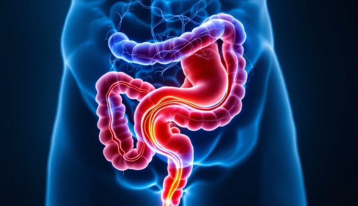
What is Enterovesical Fistula?
A fistula is an abnormal link between two tissue surfaces covered by a protective layer known as the epithelium. However, some exceptions to this definition occur. For example, a fistula could be between two blood vessels, which are covered by a different kind of cell layer called endothelium. A fistula can also form from the lining of the gastrointestinal tract (our digestive system) to a wound where no protective tissue layer is present.
One type of fistula, known as an enterovesical fistula, is where an abnormal connection forms between the intestine and the bladder. The organ where the fistula starts from usually gets named first. In this case, “entero” refers to the intestine and “vesical” refers to the bladder, indicating that the fistula starts at the intestine and ends at the bladder. However, sometimes the fistula could start from the bladder and end in the intestine or other tube-like structures. Most fistulas that doctors see in their practice start from the bowel.
Now, the term “bowel” is often used to refer to the small intestine. However, in medical literature, it can be used to refer to any fistula between all parts of the intestine (both small and large) and the urinary bladder. More specific terms like jejunovesical (from the jejunum, a part of the small intestine), ileovesical (from the ileum, another part of the small intestine), colovesical (from the colon), sigmoid vesical (from the sigmoid colon, the lower part of the colon), or rectovesical (from the rectum, the final part of the colon) are used to indicate exactly which part of the intestine the fistula is coming from. The most common type of fistula between the intestine and bladder is the colovesical fistula. Most of the information you’ll find online about intestines-bladder fistulas will refer to this type, unless it’s specified otherwise.
What Causes Enterovesical Fistula?
An enterovesical fistula is a problem that can happen because of a disease or injury. It’s important to understand how this fistula, or abnormal connection, forms. This will help in treating and preventing it. This problem can happen for many reasons, and depending on the cause, it takes anywhere from months to years to form. Usually, any problem with the wall of the bowel or bladder can lead to fistula formation. Other causes include things like injury, including injuries caused during medical treatment or by radiation.
The most common reasons for an enterovesical fistula are:
1. Diverticular disease, which is when small bulging pouches develop in the digestive tract, is the most common cause. It is responsible for two-thirds or more of these fistulas. This disease is much more common in the large bowel (colon) than the small bowel (small intestine). Complicated diverticulitis, which is when these pouches get infected or inflamed, is more likely to cause a fistula. Inflammation and a small collection of pus can break down the wall of the pouch and can spread to the nearby bladder wall, forming this abnormal connection. An increase in pressure in the pouch or bladder can keep this fistula open.
2. Cancerous growths are the second main cause of enterovesical fistula. They account for about 10% to 20% of the cases. Intestinal cancer usually spreads into the sides and circumference of the intestine. The growth and damage of normal tissues may expand to the nearby bladder wall creating the abnormal connection. Similarly, bladder cancer can cause the same problem.
3. Other causes include Crohn’s disease (a type of inflammatory bowel disease), radiation treatment, and chemotherapy, both of which can cause fistulas to form years after treatment. In rare cases, injury from pelvic surgeries or traumas to the pelvis can lead to fistula formation, as can foreign bodies inside the bladder or intestines.
It’s important to note that there are factors that can cause a fistula to not heal, but these factors don’t necessarily cause a fistula to form in the first place. For instance, a fistula that is already formed is unlikely to heal if there is epithelialization, which is the process where new skin cells form over a wound, or if the distal stream of the gastrointestinal tract, or the normal path that food and waste follow, is blocked
.
Risk Factors and Frequency for Enterovesical Fistula
Colovesical fistulae, which are connections between the bowel and the urinary bladder, are the most common type of such connections. They occur in about 2% of patients with diverticular disease and less than 1% of colon cancer patients.
These fistulae are more frequently found in males due to some female-only factors. For example, in females, the uterus and adnexa lie between the bladder and the colon, preventing the formation of colovesical fistulae. Consequently, other types of fistulae are more common in females. Females who do have colovesical fistulae, are generally older or have a history of hysterectomy, and it can be suspected that the reduced size or absence of the uterus could be contributing factors.
- Colovesical fistulae, connections between the bowel and urinary bladder, are very common.
- They affect about 2% of patients with diverticular disease and less than 1% of colon cancer patients.
- These fistulae occur more often in males.
- In females, other factors, such as the position of the uterus and adnexa, make other types of fistulae more common.
- Typically, older females or those who have had a hysterectomy are more likely to develop colovesical fistulae.
Signs and Symptoms of Enterovesical Fistula
Enterovesical fistula, a disease often discovered through surgical procedures, can cause several signs and symptoms. These can be related to the cause of the disease, the disease itself, and its complications. Sometimes, the fistula could be the first sign of the underlying disease. The most recurrent signs and symptoms of this disease include repeated urinary tract infections and pneumaturia, which is air bubbles in the urine.
The signs and symptoms can usually indicate the cause of the disease:
- In case of Diverticular disease, you may experience pain along with symptoms of a gastrointestinal infection such as weight loss, weakness, poor appetite and also symptoms related to digestive problems like bowel obstruction, bleeding, change of habit and abdominal pain.
- If the cause is an Inflammatory process, then common symptoms include abdominal pain, tenderness in the lower belly, fever, gastrointestinal bleeding, and alteration of bowel habits.
If you have a known medical history related to illnesses that can cause enterovesical fistula, it should be a hint to the potential issue.
In the disease itself, a symptom that strongly suggests enterovesical fistula is pneumaturia which is when air bubbles appear in urine. Despite its rarity, the presence of stool in the urine, a condition known as fecaluria, is also a potential sign of this disease.
In terms of complications from enterovesical fistula, frequent or continuous urinary tract infections are the most common.
Testing for Enterovesical Fistula
In the case of suspected enterovesical fistulas, which are abnormal connections between your intestine and bladder, several steps are taken. These include confirming if you have the condition, investigating the location, size, and complexity of the abnormal connection, identifying the underlying cause if it’s unknown, planning for treatment, and keeping track of how the situation is progressing.
There are several ways doctors assess this condition. Normally, they start with simpler tests, moving on to more complicated ones based on necessity. Diagnosis is usually achieved through medical imaging.
As part of this assessment, you’ll undergo a thorough clinical evaluation that includes a detailed discussion about your medical history and a physical exam. There are several approaches doctors can take to assess enterovesical fistulas.
In terms of imaging, a procedure where a contrast substance that can be seen on an X-ray is passed through your gastrointestinal tract to your bladder can help confirm the diagnosis. Sometimes, the contrast may not show up in the fistula (the abnormal connection) but can be seen in the end organ, which is the bladder.
Procedures like a small bowel follow-through or contrast enema can be used to confirm the diagnosis. Computed Tomography (CT) scans can be used to provide more details about the tissues in the area and the fistula. This is useful when planning for any surgical intervention.
Magnetic Resonance Imaging (MRI) may be needed if the fistula is difficult to diagnose. MRI is good at defining the characteristics of soft tissue and can be useful in complicated cases, like in those with Crohn’s disease, an inflammatory bowel disease that can lead to fistulas.
Endoscopy is another helpful tool in diagnosing enterovesical fistulas. This is a procedure where a flexible tube with a camera on the end is used to look inside your digestive tract or bladder. If a fistula is present, the doctor could see a small area of red and inflamed tissue, indicating the location of the fistula. However, the inside of the fistula is usually difficult to see, unless it is very wide.
Endoscopy can also provide information about the underlying disease that could have caused the fistula, like cancer or Crohn’s disease. Sometimes, fistulas can be discovered incidentally during an endoscopy that’s conducted for other reasons. In such cases, further tests will be needed.
Treatment Options for Enterovesical Fistula
An enterovesical fistula is a communication between the bladder and the intestines, which can cause urinary tract infections (UTI) and other complications. The treatment of an enterovesical fistula generally involves addressing the fistula itself and treating any underlying disease that may have caused it. Therefore, a concrete diagnosis of the fistula’s cause is essential for planning an appropriate treatment strategy.
Sometimes, less aggressive or non-surgical treatments can be used. This might be suitable for patients at high risk for surgical complications or those with severe underlying diseases. Medical treatment might involve managing any UTIs, treating symptoms, and optimizing treatment for any underlying diseases like Crohn’s disease or diverticulitis. Some patients may also improve with support of their overall health.
Other non-surgical treatments can include using substances like fibrin glue to close the fistula, although the success rate for these measures is not as high as with surgical treatments. Nonetheless, they may be worth considering in higher-risk patients.
When it comes to the surgical approach, the typical method is to remove the part of the bowel involved with the fistula. After diagnosing the fistula and identifying the underlying disease, the surgeon can then appropriately plan the surgery. A conservative excision of the affected bowel and the fistula is often recommended for cases related to diverticular disease, limited Crohn’s disease, and similar reversible inflammatory diseases. In these cases, the bladder can usually be closed with an absorbable stitch, and a urinary catheter may be used for a few weeks to help with healing.
For cancerous fistulas, a more extensive excision is often necessary. This can involve removing part of the intestine and a portion of the bladder that includes the fistula.
During surgery for the underlying disease, an enterovesical fistula may sometimes be discovered. The suspicion might be triggered by unusually firm or ‘adherent’ tissue between the intestine and the bladder. In such cases, unless it is cancer surgery, the surgical approach typically remains the same. However, if cancer isn’t yet confirmed, a sample of the fistula tissue may be taken for rapid examination in the lab to rule out malignancy.
What else can Enterovesical Fistula be?
The following conditions could be responsible for certain symptoms and should be considered as part of the diagnosis:
- Aortitis
- Appendicitis
- Blunt abdominal trauma
- Colon cancer
- Diverticulitis
- Enterovesical fistula
- Inflammatory bowel disease
- Malabsorption
- Peptic ulcer disease
- Penetrating abdominal trauma
- Small intestinal diverticulosis
- Urinary tract infection


