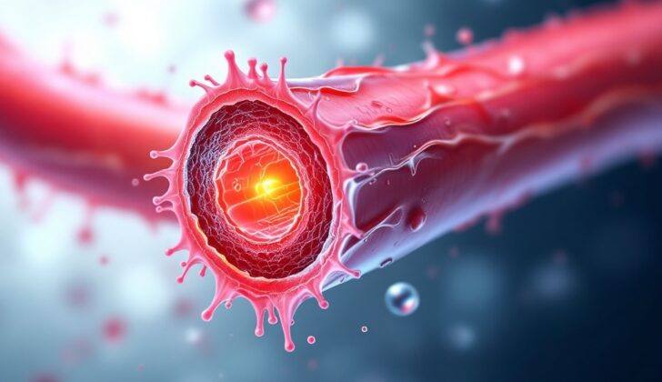
What is Neointimal Hyperplasia?
Atherosclerosis, a condition where arteries harden due to fatty deposits, is a major cause of sickness and death worldwide. To manage this, doctors perform procedures like balloon angioplasty (inserting and inflating a small balloon inside the blocked artery), with or without inserting a metal scaffold (stent), endarterectomy (surgical removal of arterial blockages), or bypass grafting (creating a new pathway for blood flow using a graft).
However, these procedures can sometimes fail due to a condition called neointimal hyperplasia (NIH). In healthy blood vessels, smooth muscle cells (SMC), which make up the middle layer (tunica media), are usually dormant, not multiplying much, and help vessels contract.
Neointimal hyperplasia is when, after surgery, these SMC start growing and moving into the inner vessel layer (tunica intima). This causes the blood vessel wall to thicken and narrows the passage for blood flow, potentially leading to the return of previous symptoms. The SMC in this condition stop helping vessels contract and instead secrete various substances that contribute to the thickening process.
Several surgical procedures can cause vessel injury triggering inflammation and recruitment of cells, which in turn can result in neointimal hyperplasia. This is often observed when a simple balloon angioplasty is done without inserting a stent or when a bare-metal stent is used. Metal stents are used to resist the vessel’s natural tendency to shrink back after angioplasty, but in the long run, they can provoke inflammation and excessive growth of the vessel’s inner lining. Also, manipulating vessels in other ways can lead to neointimal hyperplasia.
Neointimal hyperplasia can occur in two primary ways:
1. Arterial neointimal hyperplasia, which is a result of manipulating arteries during endarterectomy or angioplasty.
2. Venous graft-associated neointimal hyperplasia, that happens due to bypass grafting or the creation of an arteriovenous graft/fistula (a connection between an artery and a vein).
What Causes Neointimal Hyperplasia?
Injury to the blood vessel wall can happen during certain medical procedures. These can include:
– The use of a special tube with a balloon on the end (known as an angioplasty catheter) that is inflated during a procedure called an angioplasty.
– The use of a tube designed to remove blood clots, called an embolectomy catheter.
– A surgery called vascular endarterectomy, in which fatty deposits are scraped from a blocked artery.
– A procedure known as arteriovenous fistula creation, where small target veins are used to create a direct connection between an artery and a vein usually in your arm, for dialysis.
– Heart surgeries like vein grafting or artery bypass surgery (technically known as coronary vein grafting or coronary artery bypass grafting), in which a vein or artery from another part of your body is used to flow blood around a blocked coronary artery in the heart.
Risk Factors and Frequency for Neointimal Hyperplasia
When a venous graft is used during a CABG (coronary artery bypass grafting), it remains open and functional in 80% of cases after one year and 60% after five years. A type of this graft, called a saphenous venous graft (SVG), is more susceptible to thickening and narrowing of the inner lining than a graft from the internal mammary artery. But, any closing of the SVG graft after five years is almost always due to new atherosclerosis, plaque buildup in the arteries.
An arteriovenous (AV) fistula or graft is used as a way to get vascular access for hemodialysis in patients with end-stage renal disease (ESRD) on dialysis. The failure rate of these AV fistulas or grafts varies from 30% to 60% at one year. Research points out that the most common issue leading to failure in 30% to 60% of these AV grafts or fistulas is neointimal hyperplasia, a thickening of the artery wall, followed by blood clots in the vessels.
Signs and Symptoms of Neointimal Hyperplasia
Neointimal hyperplasia is a medical condition that can cause a recurrence of troubling symptoms. Some of these may include:
- Anginal symptoms or acute coronary syndrome for individuals who have had a coronary artery venous graft or stenting. This is particularly notable when there is critical narrowing (stenosis) of the graft.
- Failure of an arteriovenous graft or fistula. This primarily affects those with end-stage renal disease (ESRD) who are using hemodialysis. It might result in complications with vascular access and ultimately delay hemodialysis treatments.
- Restenosis, or the re-narrowing of the carotid artery after a carotid endarterectomy (CEA) due to neointimal hyperplasia. This can present as a transient ischemic attack (TIA) which is like a mini-stroke, or with stroke symptoms.
Testing for Neointimal Hyperplasia
If your doctor is looking into your health condition, they may recommend a few tests, here’s what they might include:
They might ask for an angiography. Angiography is a medical imaging technique that allows doctors to visualize the inside of your blood vessels. This helps them to see if there’s any blockage or abnormality in your blood flow.
Intravascular ultrasonography is another test that they might recommend. It’s an ultrasound performed from inside the blood vessels. It provides detailed and accurate images of the walls of the blood vessels, this helps doctors understand more about possible blockages or abnormalities.
They might also suggest intravascular optical coherence tomography. This type of imaging gives very high-resolution pictures of the inside of your blood vessels. It’s often used to study a condition called neointimal hyperplasia, which is the buildup of cells inside your blood vessels. This build-up can lead to narrowing of the blood vessels making it harder for the blood to flow.
Your doctor could also use flow Doppler catheters. These are special devices that use sound waves to measure the speed and direction of your blood flow. They provide useful information about potential flow limitations.
Graft Surveillance might be suggested too. If you’ve had a vascular graft surgery, the graft that was placed might also develop neointimal hyperplasia. So, your doctor will regularly check the graft to detect any early signs of this condition, and fix it if needed.
Lastly, they might use Carotid Doppler ultrasonography. This is an imaging method that uses high-frequency sound waves to produce images of the carotid arteries in your neck. It helps the doctors understand the velocity and direction of blow flow in these arteries. This can indicate possible issues like blockages.
Treatment Options for Neointimal Hyperplasia
Neointimal hyperplasia, which is the thickening of the blood vessel wall, can potentially be prevented and treated with medications aimed at specific stages of the condition’s development. Monitoring patients who are at high risk, particularly within the first two years after a procedure to fix their blood vessels, can help catch and treat this condition early on.
Different types of medications can be used to tackle different steps of this disease. For example, medications such as aspirin, clopidogrel, abciximab, heparin, or hirudin can be used to prevent unnecessary clot formation and platelet activation, both of which can lead to neointimal hyperplasia. A drug called probucol, which is an antioxidant, can also help prevent the condition by blocking white blood cells from entering the damaged parts of a blood vessel wall.
The lack of nitric oxide, a molecule that helps keep blood vessels healthy and functioning properly, can lead to the development of neointimal hyperplasia. Local treatments such as nitric oxide-based therapies and certain types of vascular wraps have been found to be better than systemic treatments, which can sometimes have unwanted side effects. Drug-eluting stents, which are tubes placed in blood vessels that slowly release medication, can also help in blocking cells from moving and duplicating, preventing the thickening of the vessel wall. These stents, which contain drugs such as paclitaxel or everolimus, have been used in cases of blood vessels narrowing again after a procedure to clear them, as well as during a procedure to open up narrowed or blocked coronary arteries.
Aside from these, gene therapy has also been studied as a potential approach. This involves modifying certain genes to target cell movement, duplication, and nitric oxide production. Beta vascular brachytherapy, a process where a small amount of radioactivity is placed inside a blood vessel to prevent cells from growing too much, has also shown to reduce thickening of the blood vessel wall a few months after application.
What else can Neointimal Hyperplasia be?
When determining the cause of a patient’s condition, the presence of neointimal hyperplasia can seem similar to a few other health problems. These should be taken into account:
- Neoatherosclerosis
- Late stent thrombosis
This means that when doctors find neointimal hyperplasia, they should also consider and test for these other conditions.
What to expect with Neointimal Hyperplasia
Understanding the nature or characteristics of the new tissue layer formed in blood vessels can help forecast long-term health outcomes. There’s data suggesting that irregularly formed tissue layers may be connected with poor long-term health outlooks.
Possible Complications When Diagnosed with Neointimal Hyperplasia
When blood vessels are not working properly, it may affect different organs in the body, leading to various health problems. These include:
- A recurrence of chest pain known as angina, or a heart attack.
- A failure of an artificially made blood vessel connection (known as an AV fistula) or a graft, which could cause delays to kidney dialysis treatments because of lost vascular access.
- Temporary disruptions of blood flow to the brain (transient ischemic attacks) or strokes due to narrowing or blood clots in the artery after surgery on the carotid artery.
Preventing Neointimal Hyperplasia
The best way to enhance a patient’s health condition is to prevent the disease from developing in the first place. This involves educating the patient about the positive impacts of regular exercise, quitting smoking, consuming a balanced diet, and avoiding a lifestyle that involves minimal physical activity.


