What is Right Aortic Arches?
Right aortic arch anomalies are rare and only occur in about 0.01 to 0.1% of the general population. These are abnormalities related to the branching and orientation of the aorta, the main blood vessel that supplies oxygenated blood to our body. These anomalies are often linked to a variety of heart defects present at birth, such as tetralogy of Fallot and truncus arteriosus, and chromosome abnormalities like DiGeorge syndrome (22q11 deletion).
Notably, a right aortic arch with mirror image branching of the vessels to the head and neck does not cause any physical heart-related issues. However, a right aortic arch can come along with other heart defects that are present at birth. In some cases, right aortic arches with certain unique branching patterns to the head and neck vessels can be associated with a vascular ring, a condition where the blood vessels form a ring around the windpipe and esophagus.
There are certain situations where the structural details of a right aortic arch matter clinically, which means they have a major impact on the individual’s health or treatment.
What Causes Right Aortic Arches?
In many people, there’s no identified genetic problem associated with the presence of a right aortic arch – which is a rare condition where the aorta, the main blood vessel in the heart, bends to the right instead of the left. However, a right aortic arch can be linked with a condition called DiGeorge syndrome (where a small part of chromosome 22 is missing) and certain birth defects of the heart such as tetralogy of Fallot, truncus arteriosus, and d-transposition of the great arteries.
In medical literature, there are generally three types of aortic arches, classified by a system known as the Edwards classification scheme. These types are:
1. Mirror image branching of the vessels supplying the head and neck. The first branch is the innominate artery, the second the right carotid, and the third the right subclavian. This is the most common and represents 59% of the cases. It’s often linked with heart diseases that are present from birth – truncus arteriosus and tetralogy of Fallot.
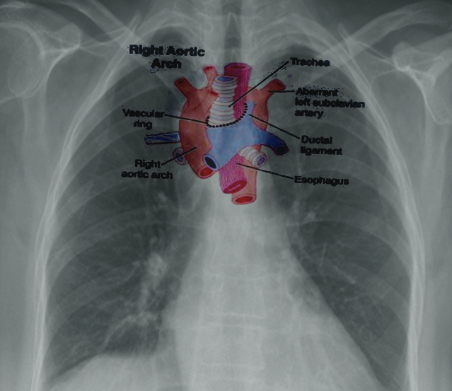
2. Aberrant left subclavian artery from a diverticulum of Kommerell as a fourth branch from the aorta. The first branch is left carotid, second right carotid, and third right carotid. This is second most common, accounting for about 40% of cases, and not usually associated with congenital heart disease. It completes a vascular ring.
3. The left subclavian artery gets eliminated or secluded with natural bypassing routes. Otherwise, the branching of the aorta is similar to type 2. This is the least common, making up about 1.5% of cases.
Risk Factors and Frequency for Right Aortic Arches
A right aortic arch is a condition where the aortic arch, a major artery in the heart, goes over the right bronchus instead of the left one. This deviation happens during embryonic development when the right side of the dorsal aorta prevails, while the left side diminishes.
Depending on how the adjacent branches of the aortic arch are formed, different types of right aortic arch can develop. This includes types like:
- Mirror-type right aortic arch: In this type, the first branch coming from the aorta is the left innominate artery. This is followed by the right common carotid artery, and then the right subclavian artery. Essentially, it mirrors the usual structure but with a right aortic arch. In this scenario, patients can be prone to left pulmonary artery stenosis or even isolation.
- Type 2 right aortic arch: The initial branch coming from the aorta is the left common carotid artery. It’s then followed by the left common carotid artery, the right subclavian artery, and finally, the left subclavian artery. If there is an aberrant left subclavian artery, it can lead to conditions such as an outpouching of the aorta or a vascular ring around the esophagus, causing it to narrow.
- There is also an extremely rare variant of right aortic arch where the left subclavian artery is isolated from a duct that’s attached to the left pulmonary artery.
- Another unusual case involves a double aortic arch with a nonfunctional left segment that can be mistaken for a right aortic arch. This differentiation is essential because symptoms and surgical treatments for these conditions will differ.
Signs and Symptoms of Right Aortic Arches
When a person has a congenital heart defect such as tetralogy of Fallot or truncus arteriosus, certain abnormal signs will show up in their medical history and physical exam. However, if there is no such heart defect, the heart examination for a patient with a right aortic arch condition would be normal. There are some specific symptoms associated with a vascular ring, another heart-related condition. These can include recurrent episodes of croup (a respiratory infection often seen in children) and a stridor (a harsh or high-pitched respiratory sound), often more noticeable in cases of double aortic arch vascular rings. Problems with swallowing, or dysphagia, are more frequently found in cases of right aortic arch and aberrant left subclavian form of a vascular ring. In some cases, referred to as “loose vascular rings,” a person may not show any symptoms throughout their entire life.
Testing for Right Aortic Arches
A chest x-ray might reveal that the windpipe, known as the trachea, has shifted to the left if a right aortic arch (a condition where the large blood vessel branching off the heart curves to the right instead of the left) is present. A common result of a left-sided aortic arch is the trachea shifting to the right, but in a right aortic arch, it’s the opposite.
A barium swallow is an examination where you swallow a liquid containing barium to highlight your esophagus on an x-ray. This could show an indentation on the back of the esophagus caused by a vascular ring (when blood vessels form an abnormal ring around the esophagus and windpipe), if present.
An echocardiogram, which is an ultrasound of your heart, will show if there are any abnormalities inside the heart. It can also reveal which side the aortic arch is on and provide detailed images of its structure. The aortic arch is identified as being on the right side if, when viewed with echocardiography, the first branch of the aorta splits to the left.
If there’s a suspicion of a vascular ring causing significant symptoms, a CT scan (a special type of x-ray) can be performed. This may be helpful to surgeons preparing for an operation, but it should only be used in patients who may require surgery for a vascular ring. CT scans can also show the condition of the airway and esophagus and can detail any squeezing or narrowing of these structures.
An MRI (a type of scan that uses powerful magnets and radio waves) of the aorta can be done instead of a CT scan to avoid any potential long-term risks from radiation. Lastly, cardiac catheterization and angiography (a type of x-ray that uses a special dye to see blood vessels) can be performed to image the aortic arch. However, because this is more invasive, it is used only for patients with other congenital heart defects that require detailed information for planned heart procedures.
Treatment Options for Right Aortic Arches
Patients experiencing symptoms from a condition known as “right aortic arch with an aberrant right subclavian” or “vascular ring,” can receive surgery to alleviate these symptoms. This surgery involves dividing a connecting tissue called the ligamentum arteriosus and pulling tight a little pouch called the diverticulum of Kommerell that’s next to the esophagus. However, if the patient also has breathing trouble due to a condition known as tracheomalacia, this breathing trouble may not immediately disappear after surgery.
In cases where the branches of the right aortic arch mirror each other, treatment might not be needed unless there’s an important irregularity in the left lung. If the left pulmonary artery, which supplies blood to the left lung, is narrow, a less invasive procedure where a balloon or a stent (a small mesh tube that’s used to treat narrow or weak arteries) is inserted can be used to widen the artery.
If the left pulmonary artery is isolated, meaning it’s not connected as it should be, a treatment plan involving restoring the function of the artery and reattaching it can be carried out.
Surgery for treating a vascular ring is usually not linked with ongoing health issues. However, there’s a slight risk that the operation might cause injury to the recurrent laryngeal nerve, which can lead to voice cord paralysis, or injury to the thoracic duct, resulting in chylous pleural effusions, a condition where a type of body fluid called lymph collects in the space around the lungs.
What else can Right Aortic Arches be?
When it’s found that a patient has a right aortic arch, it’s crucial to understand how the aortic arch branches out. This allows doctors to inform families and patients about the likelihood of developing symptoms. As a rule of thumb, symptoms from birth defects in the heart typically appear during infancy and cause more worry than those related to a vascular ring.
Issues with feeding and swallowing in kids are usually linked to acid reflux, while respiratory symptoms, which are less likely with a right aortic arch, are commonly found in conditions like asthma, croup, or laryngomalacia.
Preventing Right Aortic Arches
Patients who have a condition called right aortic and mirror-image branching shouldn’t worry too much – this is usually harmless. This condition can sometimes create a kind of ring around the windpipe and esophagus, known as a vascular ring, but this rarely causes significant breathing issues. Typically, treatment is recommended to help improve symptoms related to swallowing.


