What is Single Ventricle?
A single ventricle or univentricular heart is the term for several different heart structure problems where one chamber, or ventricle, is not fully developed. It can also refer to a condition where a wall separating the ventricles didn’t form. Because of these abnormalities, the heart can’t separate oxygen-rich blood from oxygen-depleted blood as it should. In most cases, this heart condition happens because of certain genes passed down in families, but certain factors in a person’s environment can also help contribute to the development of these heart malformations.
What Causes Single Ventricle?
The conditions and their origins can differ vary based on the type and are affected by several factors. We don’t fully understand how each kind develops.
Abnormalities can occur while the baby is developing in the womb, specifically between day 30 to 56. It’s common for heart structure abnormalities to be linked with conditions like situs inversus totalis and heterotaxy, which involve the organs being in the wrong place or being arranged differently. In patients with primary ciliary dyskinesia, a condition that affects the movement of the hair-like structures (cilia) on cells, 12% of them had signs of heterotaxy. We know of genetic causes too. Tbx5 and GATA4, for example, directly impact the formation of the ventricular septum, the wall separating the lower chambers of the heart. There are also cases where the cause isn’t known but could be linked with a defect in the formation of the endocardial cushions, structures that eventually form heart valves and septa, or influenced by the flow of blood. These cases are related with non-heart structural abnormalities, like those seen in DiGeorge Syndrome.
Environment also plays a role in the development of the heart structure. Some of the risk factors include:
Select univentricular heart abnormalities and their typical characteristics include:
Furthermore, an atrioventricular canal defect occurs when an opening is large enough in an atrial or ventricular septal (the wall separating the heart’s chambers) to effectively form one ventricle instead of two.
Risk Factors and Frequency for Single Ventricle
Congenital heart disease, a birth defect that affects the way the heart works, is found in roughly between 6 and 13 out of every 1,000 live births. There are several types of congenital heart diseases, and their occurrence rates vary.
- Hypoplastic left heart syndrome, the most common type of congenital heart disease that affects just one chamber of the heart, is seen in 2 to 3 out of every 10,000 births, and it’s more common in males.
- Tricuspid atresia, a type of heart defect where a valve in the heart fails to develop, occurs in roughly 1 out of every 10,000 live births.
- Ebstein anomaly, a rare heart defect where the tricuspid valve is misplaced in the heart, happens in about 0.5 out of every 10,000 live births. Interestingly, if a mother uses lithium during pregnancy, the occurrence of this defect can increase nearly seven times.
- Double outlet right ventricle, a condition where both major arteries connect to the right ventricle instead of one to each ventricle, occurs in 0.009 out of every 10,000 live births.
- Double inlet left ventricle, a defect where both major veins connect to the left ventricle instead of one to each ventricle, can occur in up to 0.01 out of every 10,000 live births.
- Atrioventricular canal defect, which is a large hole in the center of the heart, happens in between 0.03 to 0.04 out of every 10,000 live births.
Signs and Symptoms of Single Ventricle
A single ventricle heart defect may show up as early as the 18th week of pregnancy. This condition can be detected with various tests. Other signs of this heart defect include misplacement of the major heart arteries and abnormal blood flow in parts of the fetal heart. An ultrasound may also show this structure outside the heart, which helps in making the diagnosis.
After birth, the symptoms can vary depending on the exact structure of the heart. Usually, a heart murmur, rapid breathing, breathing problems, bluish skin, or low blood pressure may be signs of insufficient blood flow and oxygen supply. Other physical signs, like an enlarged liver or unusual physical features, can also hint at more underlying problems. A newborn may not show symptoms at birth or before being discharged from the hospital if blood flow is adequate at the time of the checkup. However, symptoms may appear after being discharged if the patent ductus arteriosus (an open blood vessel that allows blood to bypass parts of the fetal heart) closes or if blood flow to the end organs changes.
A single ventricle heart defect may show up as early as the 18th week of pregnancy. This condition can be detected with various tests. Other signs of this heart defect include misplacement of the major heart arteries and abnormal blood flow in parts of the fetal heart. An ultrasound may also show this structure outside the heart, which helps in making the diagnosis.
After birth, the symptoms can vary depending on the exact structure of the heart.
- Heart murmur
- Rapid breathing
- Breathing problems
- Bluish skin
- Low blood pressure
These may be signs of insufficient blood flow and oxygen supply. Other physical signs, like an enlarged liver or unusual physical features, can also hint at more underlying problems. A newborn may not show symptoms at birth or before being discharged from the hospital if blood flow is adequate at the time of the checkup. However, symptoms may appear after being discharged if the patent ductus arteriosus (an open blood vessel that allows blood to bypass parts of the fetal heart) closes or if blood flow to the end organs changes.
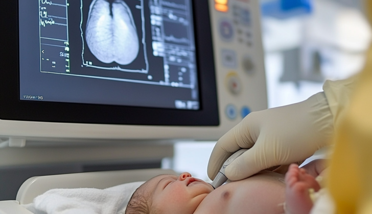
Testing for Single Ventricle
When expecting a baby, doctors use standard ultrasound techniques and special heart ultrasound (fetal echocardiography) to check the baby’s development. These tools can identify irregular heart structures or unusual patterns in the blood flow, all of which might indicate a single ventricle in the baby’s heart before birth.
After the baby is born, several methods are used to confirm whether they have a single ventricle. The most effective method is echocardiography, which is another type of heart ultrasound. Other tests such as ECG (electrocardiography), chest X-rays, and pulse oximetry (which measures how much oxygen is in the blood) can also help diagnose this condition. In addition, the doctor may find clues during a physical examination.
There are also other imaging techniques like CT scan, invasive test like cardiac catheterization, and MRI, including a special one called cardiac magnetic resonance imaging, which can show if the heart has a single ventricle. But these advanced tests are usually saved for complicated cases or when treatment needs to be planned.
Treatment Options for Single Ventricle
The treatment options for univentricular heart conditions, or situations where a person only has one functioning ventricle in their heart, need to be carefully chosen based on when the condition is discovered and the overall health outlook. It’s important to thoroughly discuss the possible outcomes before deciding on a treatment, as there may be cases where a procedure may not substantially improve the condition.
Medical treatment for univentricular heart conditions is usually focused on managing the specific disease that is causing the problem. For example, extra oxygen can be given to relieve low oxygen levels in the blood, and imbalances in the body’s pH or metabolites should be corrected. Some patients may benefit from a medication called inhaled nitric oxide, which can lower resistance in the lung’s blood vessels and allow more blood to get oxygenated by the lungs. Certain medications may assist the heart in contracting more forcefully, but others that can affect heart rate should be avoided.
Using a medicine called prostaglandin E1 can be useful if a blood vessel known as the ductus arteriosus needs to stay open to ensure blood flow. It’s also important to avoid certain medications, such as non-steroidal anti-inflammatory drugs, which can prevent the ductus arteriosus from staying open.
Catheter-based treatments, which involve inserting a tube into a blood vessel to perform procedures, are another option. Such treatments focus on the specific cause, timing, and prognosis of the condition. Interventions can be performed in the womb or after birth to correct defects. In some cases, blockages in pulmonary blood vessels can also be dealt with using this approach.
Surgical procedures are also an option for correcting these heart abnormalities. The specific technique used will depend on the patient’s particular condition. One option is the Fontan procedure, which reroutes blood to the lungs using the body’s natural pressures. However, this surgery should not be the only one considered, as it’s important to examine each case individually.
For severe cases, surgeries focused on providing comfort, rather than curing the disease, may be a more suitable choice. Sometimes, these types of surgeries can have unclear outcomes. In such cases, it might be recommended for the patient to be referred to a specialized institution for further assessment and care.
Heart transplantation can also be considered but may come with less than ideal outcomes due to other health issues. Even after other treatments, transplant may still be required.
What else can Single Ventricle be?
Babies might show signs of certain health issues right after birth or a few days post-delivery as their secondary circulations reduce. In emergency situations following birth, conditions such as sepsis, Infantile Respiratory Distress Syndrome (IRDS), independent heart and lung abnormalities, and misplacement of the heart’s main blood vessels should be suspected.
Milder cases may become apparent few months or years later when the child’s growth rate is impacted. In these instances, issues like infections, failure to gain weight and grow at the expected rate, and insufficient nutrition should be contemplated.
What to expect with Single Ventricle
Hypoplastic left heart syndrome can be fatal if left untreated, yet improves to a 60% to 70% survival rate with certain medical interventions. Evidence shows that patients who survive beyond one year after a surgical fix are 90% likely to live until 18. However, regardless of the treatment, it’s important to understand there’s a heightened risk of slower mental development. Because of this, the American Heart Association recommends routine developmental checks, explaining that it might be related to insufficient nutrition.
Patients diagnosed with tricuspid atresia generally fare better with intervention, and statistics point to a 90% survival rate at one year of age and an 80% survival rate at ten years. Ebstein anomaly, on the other hand, has a high mortality rate in newborns, with about 32% dying before hospital discharge. Their survival rates for one and ten years have been recorded as 67% and 59% respectively. Unfortunately, data about prognosis after ten years is limited.
The outlook for other causes of single ventricles varies, but generally, more than half of the individuals survive for two years, and on average, can live for 30 to 40 years.
Possible Complications When Diagnosed with Single Ventricle
People with single ventricle heart syndrome usually undergo surgical intervention to correct the problem. However, this can sometimes make it hard to pinpoint the cause of certain complications that might later arise. Below are the complications that are expected:
- Arrhythmias, which is an irregular heart rhythm
- Esophageal varices, which are enlarged blood vessels in the esophagus
- Heart failure leading to the formation of blood clots, with an increased risk of bleeding from treatment
- Increased risk of complications when undergoing anesthesia
- Long-term condition called cyanosis, where the skin, lips, and fingernails turn blue due to lack of oxygen in the bloodstream
- Respiratory diseases that make it difficult to breathe due to restriction or damage to the lung tissues. These might be partially caused by unnoticed issues, such as plastic bronchitis and unnoticed lung blood clots
- Protein-losing enteropathy, a condition that affects the body’s ability to absorb proteins from the diet
- Injury to the recurrent laryngeal nerve, which may affect voice
- Short stature and delayed physical development
- Problems with kidney function
- Formation of abnormal connections between arteries and veins in the lungs
Preventing Single Ventricle
Treatment isn’t always the best or most suitable choice. It’s crucial to have detailed conversations about the expected medical condition and what outcomes to anticipate with family members. It must be clearly explained that any staged procedures won’t necessarily cure the condition and there may be a future need for a heart transplant. In the case of unborn babies, the discussion might need to involve the option of ending the pregnancy. It’s also important to talk about the patients’ living conditions. For example, families living at heights greater than 1700 meters above sea level may not have as many long-term health benefits because of their altitude.


