What is Basilar Skull Fractures?
Basilar skull fractures are injuries caused by intense blunt force that affect at least one bone at the bottom of the skull. These fractures most often impact the bones around the ears, but they can also damage the bones at the back of the skull, as well as the bones near the nose, eyes, and forehead. Certain visible signs indicate a high possibility of a basilar skull fracture. These include blood behind the eardrum, fluid leakage from the ear or nose, discoloration behind the ears (known as Battle sign), and black and blue coloring around the eyes (known as raccoon eyes).
These fractures are often accompanied by other injuries, such as facial fractures, neck injuries, brain bleeding, nerve injuries, blood vessel damage, and meningitis. Basilar skull fractures are more common in younger individuals who engage in high-risk activities. The majority of these fractures are treated with non-surgical care.
What Causes Basilar Skull Fractures?
Most of the time, basilar skull fractures occur due to high-speed blunt force, like car accidents, motorcycle crashes, or injuries to pedestrians. Falls and assaults also contribute significantly to these fractures. Less than 10% of these fractures come from penetrating injuries, such as gunshot wounds.
Risk Factors and Frequency for Basilar Skull Fractures
Basilar skull fractures, which are breaks in the base of the skull, are not very common. They occur in roughly 4% of people who have a severe head injury. Despite this, they make up about 19% to 21% of all skull fractures.
Signs and Symptoms of Basilar Skull Fractures
Basilar skull fractures are characterized by a range of symptoms, which can vary depending on the severity of any associated brain or cranial nerve injury. People with this condition may experience changes in their mental state and could be nauseous or vomit frequently. There may also be issues with eye movement, due to damage to cranial nerves III, IV, and VI, and potential facial droop because of injury to cranial nerve VII. Problems such as hearing loss or ringing in the ears might indicate harm to cranial nerve VIII.
There are certain telltale signs linked to basilar skull fractures:
- Hemotympanum: This refers to the pooling of blood behind the eardrum, which gives it a purple appearance. This typically happens within hours of injury and could be the earliest detectable sign of a skull fracture.
- Cerebrospinal fluid (CSF) rhinorrhea or otorrhea: Known as the “Halo” sign, it describes the double ring pattern that appears when bloody fluid from the ear or nose – containing CSF – is dripped onto paper or cloth. This sign is not unique to the presence of CSF, as other liquids like saline or tears can also create a ring pattern when mixed with blood.
- Periorbital ecchymosis (raccoon eyes): This refers to blood pooling around the eyes, a sign frequently associated with fractures of the anterior cranial fossa. Usually, this symptom appears 1 to 3 days after the initial trauma and is a strong indicator of a basilar skull fracture when it occurs on both sides.
- Retroauricular or mastoid ecchymosis (Battle sign): This is characterized by the accumulation of blood behind the ears in the mastoid region, indicating fractures to the middle cranial fossa. Similar to raccoon eyes, this sign often presents itself 1 to 3 days post-injury.
In addition, people may experience dizziness, ringing in the ears (tinnitus), and uncontrolled eye movements (nystagmus).
Two clinical signs, Battle sign and raccoon eyes, are highly indicative of a basilar skull fracture.
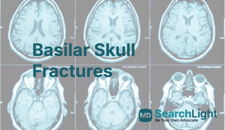
Testing for Basilar Skull Fractures
In some cases, a doctor may be able to diagnose a basilar skull fracture during a physical exam. Regular x-rays, however, may not be sensitive enough to detect this type of injury.
Usually, doctors start with a special type of scan called a non-contrast computed tomography (CT) scan. However, some specific types of fractures, particularly those that are straight or have not moved from their original position, can be challenging to spot.
In cases where the doctor strongly suspects a basilar skull fracture, a more detailed CT scan might be useful. This scan, called multidetector CT (MDCT) thin-slice scanning, produces images of the face and the base of the skull. These images can help identify more subtle fractures. However, it is also possible to mistake the detailed images of small nerve pathways and blood vessels as fractures. Breach of the cranial cavity causing air to enter it should alert the doctors about the probable presence of a basilar skull fracture.
Further imaging with CT angiography and venography (CTA, CTV), types of scans that display the blood vessels, can be utilized to check for any potential vascular injuries. In specific situations, an MRI scan can be helpful to assess whether a nerve injury has occurred or to check for leakage of cerebrospinal fluid.
Detecting a cerebrospinal fluid leak can be difficult. If leakage is suspected, the fluid should be collected and examined for beta transferrin, a protein found in cerebrospinal fluid.
Treatment Options for Basilar Skull Fractures
Basilar skull fractures are typically the result of serious injury. Immediately treating issues related to airway stability, breathing, and blood circulation becomes the top priority in these cases. Also, injuries to the cervical spine are common in these situations, so it’s crucial to immobilize the cervical spine, particularly when managing the airway. Procedures such as inserting a nasogastric tube or performing nasotracheal intubation should be avoided due to the risk of accidentally placing the tube into the brain.
Additionally, nasal intermittent positive pressure ventilation – a breathing aid that delivers pressurized air through the nose – should be avoided as it could potentially cause air accumulation in the skull.
Patients with basilar skull fractures need to be hospitalized for monitoring. If they are taking blood-thinning medications, they should be taken to a facility with immediate neurosurgical capabilities and the ability to frequently assess for any neurological deterioration, even if there’s no bleeding visible on initial imaging. Patients who have bleeding within the skull require an urgent evaluation by a neurosurgeon. Otherwise, fractures at the base of the skull are often managed with a watch-and-wait approach. Surgery could be required in complications such as intracranial bleeding needing decompression, vascular injury, significant cranial nerve injury, or ongoing leakage of cerebrospinal fluid (the fluid that surrounds the brain and spinal cord).
Basilar skull fractures heighten the risk of meningitis since bacteria from the sinuses, nasopharynx, and ear canal can make direct contact with the central nervous system. Up to 45% of patients with basilar skull fractures develop cerebrospinal fluid leaks. There’s a common practice of using preventative antibiotics for these patients to protect against meningitis, but there isn’t strong evidence to back this approach. A recent comprehensive review didn’t find enough evidence to recommend preventative antibiotics in patients with basilar skull fractures, even if they have a confirmed cerebrospinal fluid leak. Nevertheless, patients with ongoing fluid leaks should have the fluid cultured to guide antibiotic therapy, and those showing signs consistent with meningitis should be treated with antibiotics while awaiting results of the cultures. Antibiotics are generally not indicated for use as a preventative measure, although they may be appropriate for use prior to procedures like the insertion of a device to monitor intracranial pressure. Persistent fluid leaks require intervention by a neurosurgeon, with less invasive endoscopic techniques becoming more commonplace, reducing the need for open surgery.
What else can Basilar Skull Fractures be?
The primary conditions that might be confused with this one include birth defects of the skull bones and a condition known as basal encephaloceles. Both of these can lead to something called a cerebrospinal fluid (CSF) leak, which presents similarly.
What to expect with Basilar Skull Fractures
The outcome of a skull base fracture can be influenced by a variety of factors, including the presence of a corresponding dural tear and cerebrospinal fluid (CSF) leak, stability of the fracture, other associated injuries, and the initial severity of any nerve or blood vessel injuries.
Interestingly, the majority of CSF leaks that can arise due to these fractures tend to resolve on their own within 5-10 days, though some may persist for several months. Again, while meningitis is not common, itching up in less than 5% of patients, its risk can increase based on how long the CSF leak lasts.
Meanwhile, conductive hearing loss, another possible complication of skull base fractures, often resolves within a period of 7 to 21 days.
Possible Complications When Diagnosed with Basilar Skull Fractures
Possible Complications:
- Leakage of cerebrospinal fluid (CSF)
- Meningitis (Inflammation of the membranes surrounding your brain and spinal cord)
- Weakness in the cranial nerves that control functions like sight, hearing, and taste
- Loss of hearing
- Formation of blood clot in a major vein at the base of the brain (Cavernous sinus thrombosis)
- Feeling of dizziness with a sensation of spinning (Vertigo)
- Bleeding inside the brain (Intracranial hemorrhage)
- Potential death
- Loss of smell and paralysis of the facial muscles (Cranial nerve deficits)
- Vascular injuries due to basilar skull fractures resulting in blockage, abnormal connections between blood vessels, bleeding, or the formation of a false blood vessel
Preventing Basilar Skull Fractures
If a patient has a condition called CSF rhinorrhea and is being treated without surgery, they should be careful not to do things like straining or blowing their nose. Furthermore, if they start to experience symptoms like a headache or fever, it’s important for them to tell their doctor as soon as possible.


