What is Diaphyseal Femur Fracture?
Broken femurs, or thigh bones, are often seen alongside other severe injuries that can lead to serious health risks. Swift treatment and careful management can result in the best outcomes for patients. In the past, these fractures were treated with just splints, but today, inserting a rod inside the bone, known as intramedullary (IM) nailing, is a common treatment method. For children, flexible rods are used to allow for ongoing bone growth.
What Causes Diaphyseal Femur Fracture?
The femur, or the thigh bone, is made up of several areas: the head (top), neck (just below the head), intertrochanteric (the area between the two bumpy bits near the top of the femur), subtrochanteric (just below these bumps), shaft (the long, straight part), supracondylar (just above the knee), and the condylar (part that connects to the knee) regions.
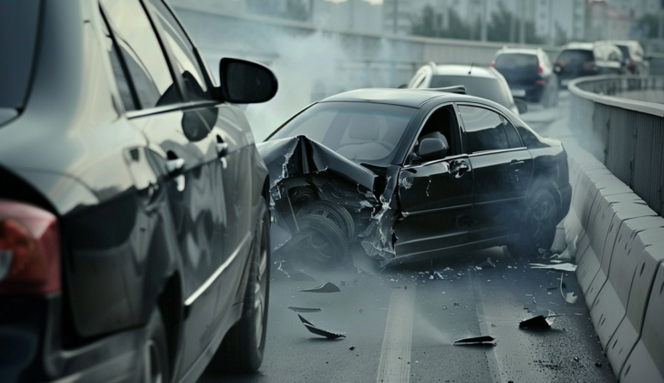
Younger people frequently break their femurs due to high-impact incidents like car accidents. Older people, on the other hand, may break this bone during a simple fall from standing height, especially if they have osteoporosis, a condition that weakens bones.
Gunshot wounds to the lower leg are another cause of femur fractures.
Stress fractures are small cracks in the bone that can occur in people who do a lot of physical activity. Lastly, some femur fractures happen due to osteoporosis or long-term use of certain medications for osteoporosis known as bisphosphonates.
Risk Factors and Frequency for Diaphyseal Femur Fracture
Femur fractures, or breaks in the thigh bone, are a fairly common problem. Each year, worldwide, there are approximately 9 to 22 fractures for every 1000 people. These injuries typically fall into two main age groups. Studies reveal that diaphyseal fractures, which are breaks in the main section of the bone, are more common in older patients. These patients often have weaker bones, a lower body mass index, and an extensive amount of bowing in the front and side of their leg.
Signs and Symptoms of Diaphyseal Femur Fracture
If a person has serious injuries, it’s crucial to check their overall condition. When they arrive at the hospital, doctors follow strict guidelines, called advanced trauma life support (ATLS), to take care of them. If the person is in a critical state or if there’s a chance they might have abdominal injuries, they could need immediate surgery. Yet, the top priority is always to save their life.
If the patient’s condition is stable, a detailed physical exam is done. Doctors may notice a visible problem with the thigh. It’s critical that they check the blood supply to the leg and see if the bone is poking through the skin.
People with fractures in both upper legs could face more lung problems and a higher chance of dying from their injuries.
Following is a summary of the points mentioned:
- Assessment of the patient’s overall condition is critical, especially in cases with potential life-threatening injuries
- Adherence to advanced trauma life support (ATLS) guidelines is paramount upon patient arrival at the hospital
- If the patient is severely ill or suspected to have internal abdominal injuries, they may need immediate surgery
- Patient’s life is always the top priority
- If the patient is stable, a thorough physical check is done, including the evaluation of the blood supply to the leg and observing for visible bone fractures
- Dual upper leg fractures could result in more lung problems and higher patient mortality rate
Testing for Diaphyseal Femur Fracture
Imaging tests, such as X-rays, are often the first step in diagnosing health issues related to your femur, which is the thigh bone. Doctors take images, or radiographs, from different directions to thoroughly assess the bone and the nearby hip and knee joints.
It’s very important to look for injuries to the femoral neck, which is located near the hip. Studies show that this type of injury happens in 1-9% of cases. To avoid missing this type of injury, many trauma centers are using CT scans. Before CT scans became common, around 20-50% of femoral neck injuries were not spotted on time.
Imaging tests are also helpful when making treatment decisions. If there’s a fracture to the diaphysis, or the main shaft of the femur, and there’s also a fracture near the upper end of the femur, a CT scan can provide crucial information. The scan can check the condition of the greater trochanter (a large, bony prominence on the upper part of the femur) and the piriformis fossa (an area located near the joint area).
Fractures to the femur can be ranked using the Gustillo-Anderson ranking system, used specifically for open fractures where the bone breaks the skin. Studies show 43% are classified as grade I, 31% grade II, and 26% grade III. Treatment for open fractures has to be aggressive due to the severity of injury and because the fracture must tear through a sizable amount of soft tissue to become open.
The Orthopaedic Trauma Association also has a classification system for femur fractures. This system covers simple fractures and also more complicated, wedge-shaped and complex fractures. The Winquist and Hansen Classification system also categorizes them, ranking them from Type 0 (no bone shattering) up to Type IV (no contact between the upper and lower fragments).
When someone fractures their femur, doctors also closely monitor their bloodwork to ensure their hemoglobin levels are healthy. Because femur fractures can cause significant blood loss, this could potentially lead to low blood pressure. In the case of an open fracture, a sample is also taken from the wound to check for infection.
Treatment Options for Diaphyseal Femur Fracture
In the case of open fractures, where the bone has broken through the skin, swift action is needed. Doctors typically give patients antibiotics – cefazolin is a common choice – and clean and debride (remove damaged tissue from) the injury straight away. Ideally, this initial clean-up operation should be carried out within the first two hours of the patient’s arrival at the hospital.
In rare cases where a person has broken both their femur (thigh bone) and the neck of their femur, it’s recommended that the neck fracture be dealt with first. This reduces the risk of complications like improper healing or blood supply problems that could damage the round, ball-like part of the bone (the femoral head). Once the femoral neck is fixed, the surgeon can then attend to the femur fracture.
One method used to manage the pain and help the surgeon keep the femur at its proper length is a form of treatment called traction. Immediately after injury, the powerful muscles in the thigh tend to contract and shorten the femur. By inserting a pin into the lower part of the femur or upper part of the tibia (shin bone) under local anesthesia, and then applying weight along the length of the leg, this can be overcome. The exact weight applied varies depending on the patient’s weight and muscle tone and provides relief once the thigh muscles have tired out.
In certain high-risk cases, such as unstable patients or those needing vascular repair (repairs to their blood vessels), doctors might choose external fixation as a temporary solution, stabilizing the fracture with metal rods inserted into the bone. Once the patient is stable, they then move to a more definitive treatment.
The go-to treatment for fractures in the middle portion of the femur (shaft/diaphyseal fractures) is a procedure called intramedullary (IM) nailing. This provides stability and allows the bone to heal naturally.
Intramedullary nailing can be performed in one of two ways; either antegrade, where the nail is started at the top of the bone, or retrograde, starting at the bottom. Both methods have been found to have similar outcomes. Antegrade nailing provides two points of entry to the bone—via the greater trochanter or the piriformis fossa. The optimal entry point depends on patient factors such as size and muscle status.
In terms of the actual nail used, it needs to match the curvature of the patient’s femur for a successful procedure. After the procedure, patients are generally advised to put weight on the leg as tolerated.
Another technique, submuscular plating, is used for special cases like complex fractures or those around a prosthetic. In this case, a plate is fixed to the outside of the bone and often necessitates limited weight-bearing after the procedure.
Lastly, research suggests operating on femur fractures within 2-12 hours after injury — as long as the patient is stable. Immediate treatment can significantly reduce complications, lower death rates, and shorten stays in the intensive care unit.
What else can Diaphyseal Femur Fracture be?
When a patient has a broken thigh bone (femoral shaft fracture), it’s important to check if they also have a fracture in the neck of the same thigh bone (ipsilateral femoral neck fracture). Depending on how they were injured, they might have other injuries too. If the injury was caused by a high-energy event like a car accident, it’s critical to rule out compartment syndrome, which is a serious condition that involves increased pressure in a muscle compartment. It can lead to muscle and nerve damage and problems with blood flow. On the other hand, if the injury happened due to a low-energy event, the fracture may be due to an underlying illness such as a tumor or a metabolic imbalance. In these cases, a thorough medical examination is needed.
What to expect with Diaphyseal Femur Fracture
The outcome for patients with a fracture in the central part of the thigh bone, or femur, depends on several factors. These include age, other health issues, whether the patient has weak or brittle bones (osteoporosis), and the treatment given. While treatment using an internal rod (IM nailing) often has good results, it’s fairly common for patients to need these rods removed later on due to discomfort.
It’s sadly not unusual for older patients to pass away after such surgery. Other complications can include infection, blood loss, and issues with the bone healing poorly or not at all, leading to potential repeat surgeries. Treatments using an external frame for support (external fixation) can be effective, but also sometimes lead to issues with infection and the bone healing at an incorrect angle.
Patients often need to stay in the hospital for a while, followed by an intense round of physical therapy. Unfortunately, many patients continue to face problems with walking and chronic pain.
Possible Complications When Diagnosed with Diaphyseal Femur Fracture
Non-unions, which are rare cases where bones fail to heal after fractures, do occur. In such scenarios, it’s vital to determine the root cause of the non-union. Sometimes, to treat this issue, a repeat surgery might be needed to improve some specific aspect of the fracture fixation, like stability or biology.
If the non-union is hypertrophic and aseptic, meaning it’s enlarged and not infected, it can be treated with compression and exchange nailing, a surgical procedure. For atrophic non-unions, where the most common symptom is a halt in the healing process, it’s important to rule out infections, especially when the fractures were previously open. The patient’s nutritional status also ought to be checked through lab tests. In treating atrophic non-unions, bone grafting, a procedure where new bone or a replacement material is placed into the spaces between or around broken bone, is usually added.
Deaths related to deep vein thrombosis (blood clots in the leg), pulmonary embolism (which happens when a blood vessel in your lungs is blocked usually by blood clots), infections, nerve injuries, and compartment syndrome (a painful condition caused by pressure buildup from internal bleeding or swelling of tissues) are not uncommon.
- Deep vein thrombosis
- Pulmonary embolism
- Infections
- Nerve injuries
- Compartment syndrome
Preventing Diaphyseal Femur Fracture
Patients who have suffered from fractures to the main bone in their thigh, known as femoral shaft fractures, need to have their healing progress closely monitored. Though it’s not common, some people can experience what are called ‘insufficiency fractures’ in this area. This type of fracture occurs when the bone isn’t strong enough to handle normal activities. However, it’s possible to prevent these fragility fractures through proper screening and medical supervision. This is particularly beneficial for those at risk, such as older individuals or those lacking in vitamin D.


