What is Distal Femur Fractures?
Fractures of the distal femur, the lower part of the thigh bone, are somewhat common. These fractures usually occur in the area above and between the knee joint. The aim of treating these injuries is to return the broken bone and joint surfaces to their correct positions and restore the limb’s proper alignment, length, and rotation. However, despite advancements in medical devices, managing these types of fractures can be difficult. This difficulty arises because the fractures often break into many pieces, affect the joint, and occur in weak, porous bones, making it tricky to secure the broken pieces.
In older adults who sustain these fractures, managing the injury can be more complex due to other health conditions they may have. These conditions can affect the treatment options available for these fractures.
What Causes Distal Femur Fractures?
Fractures in the lower part of the thigh bone, or the ‘distal femur’, affect adults in two main ways. For young men, these fractures are usually the result of high-energy events like car accidents. However, for older people, these fractures often occur due to low-impact incidents like falls from standing height.
It’s important to note that older patients often suffer from other serious health conditions. These can hinder their ability to undergo surgery, affect their recovery, and even influence their chances of survival.
In children, the real issue often lies in the long-term effects of these injuries. If fractures near the joints aren’t treated correctly, they can cause early joint damage. As our population grows older, treating these complex fractures becomes a challenge and often ends up having less than ideal results.
Risk Factors and Frequency for Distal Femur Fractures
Distal femur fractures are relatively rare, comprising less than 1% of all fractures and about 3 to 6% of fractures in the femur, the large bone in the thigh. These types of fractures are seen in two main groups: younger men who have experienced a serious accident involving a vehicle, and older women. It’s noted in one study that in patients 35 or older, who suffered this type of fracture due to moderate trauma, 80% of them showed signs of generalized bone thinning or osteopenia. Also, fractures near artificial knee joints have become more common. The occurrence of distal femur fractures around an initial total knee replacement ranges from 0.3% to 5.5%, and this jumps to as high as 30% in cases where the knee replacement had to be redone.
Signs and Symptoms of Distal Femur Fractures
If a person is injured by a high energy incident, they will need to go through a complete trauma assessment, following the advanced trauma life support protocol. Some key signs of this type of injury include severe pain in the thigh and knee area, and the inability to put any weight on the injured leg. Sometimes, there can be a noticeable unusual shape or bending in the lower thigh and knee. If you suspect that the injury is an open fracture (a type of injury that affects 5-10% of certain types of fractures), the skin around the area needs to be examined closely.
- Immediate tetanus prevention and intravenous antibiotics should be given following an open fracture.
- Make sure to clean the wound and remove any dirt or foreign objects promptly to avoid infection.
The blood flow and nerve functions of the affected leg need to be checked before and after any adjustments are made to the bone position. If there’s any difference in pulse strength between the injured and non-injured legs, or if the blood flow isn’t normal, more tests need to be done. If there is a suspicion of damage to the blood vessels, a CT scan with a consultation from a vascular surgeon will be needed.
The treatment approach can vary depending on the patient’s age and the nature of the injury. For instance, elderly patients who have had a minor fall may not require emergency treatment; however, these types of fractures can often be associated with other injuries in younger adults because they usually occur due to high-impact damage. Importantly, an athlete who is an adolescent might have a non-displaced fracture in the lower part of the thigh bone (femur), which could easily be overlooked, and may also have other knee injuries.
Testing for Distal Femur Fractures
When a patient is suspected of having a bone injury, the doctor might take images, or radiographs, of the whole limb. This includes the joints above and below where they think the injury might be. These images help the doctor to understand the nature of the injury. Fractures of the femur, which is the thigh bone, are rarely life-threatening, but they can look alarming. Sometimes, your doctor might decide to take views of your radiographs while applying tension to your leg. This can be painful, so pain relief would be provided before your leg is manipulated. Sometimes your doctor may decide to take images of your other femur to make plans for surgery.
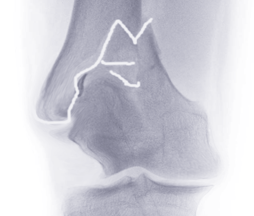
When the doctor is planning the treatment, they may decide to use a technique known as CT scanning. This is recommended for complex fractures or when there’s extension into the joints along with osteochondral fragments inside the joint space. CT is very useful for looking at a particular type of fracture, called a Hoffa fracture. This type of fracture happens at the end of the femur, and more than 38% of them involve the outer part of the knee joint. If your treatment involves an external fixture to hold your bones in place whilst they heal, a CT scan should be done after it’s fitted.
A CT angiography, a method to see the blood vessels, might be needed if your pulses lower down the limb are weak after resetting the bone, or if the pressures in your arteries are too low. These kinds of scans are especially important if the knee joint is dislocated. A specific type of CT angiography, known as a multi-slice helical CT angiography, is very good for assessing and managing injuries to the lower part of the limb.
Distal femur fractures, which are fractures at the very end of the femur, are classified in several ways, such as whether they involve the joint surface or not, and the number of fragments and how fragmented they are. The Orthopedic Trauma Association Classification is most commonly used to classify these fractures. The fractures can be just in the bone (extra-articular), partially in the joint (partial articular), or completely involve the joint (complete articular). Each one is further classified by the nature of the fracture.
Treatment Options for Distal Femur Fractures
When treating fractures that don’t affect the joint and are stable and slightly displaced, a non-surgical approach is often utilized. In these cases, patients wear a knee brace designed to encourage a range of motion to prevent stiffness, and they’re typically advised not to put any weight on the leg for six weeks. For bedridden patients with severe health conditions that make surgery risky, the fracture may be managed with splints, braces, or traction. However, these methods may cause complications such as bed sores, blood clotting, and severe knee dysfunction. Therefore, surgery can still be beneficial even for bedridden patients to avoid these complications.
All suitable patients should be offered medication to lower their chances of developing a blood clot. This is especially important for elderly patients who are at high risk. Medical practitioners should discuss the potential risks and outcomes with their patients as soon as possible, including the possibility of death or the loss of independence. Pain control through surgical interventions is not something that can prevent symptom relief efforts.
External fixation can be a temporary measure to maintain the length, alignment, and rotation of the limb until the soft tissue is ready for internal fixation. This method is used for patients with open wounds, poor skin healing, or who are too unstable for internal surgery. Complications may include infections, reduction loss, malformation, and knee stiffness.
Open reduction internal fixation is another option that includes using devices like fixed angled blade plates, sliding barrel condylar plates, condylar buttress plates, and locking plates. The main goal is to restore the normal structure of the joint surface and limb while maintaining blood circulation. The choice of surgical exposure and implant is usually done based on the fracture type and surgeon preference.
Accurate fracture reduction is very important for a successful outcome. This can be achieved indirectly, which is beneficial as it minimally invades soft tissue, thus improving fracture healing. Various devices can be used for this process, including manual traction and forceps. A combination of direct and indirect methods can also be effective.
For certain types of fractures, a chisel and plate often used can result in condylar misplacement if not correctly inserted. These devices allow compression over femoral condyles and can work better in patients with soft bones due to only needing correction in two planes compared to blade plates. However, this type of device may cause side effects due to its bulky design, and may cause joint pain and symptoms.
Condylar buttress plating and locking plates are beneficial to treat fractures of the condylar type. Locking plates have become more commonly used for fixation because of their stability and efficiency. However, potential complications could include high-energy fractures, poor reduction of the fracture, poor plate positioning, and weight-bearing before proper healing.
Intramedullary nails can provide a stable structure with minimal invasion of soft tissues and offer antegrade and retrograde options depending on the fracture. These nails allow the treatment of fractures on the same side of the hip and other fractures in severely injured patients. Potential disadvantages could include knee infection and pain, and metallosis from nail or screw breakage.
Bone cement can be beneficial in treating severely osteoporotic supracondylar distal femur fractures while bone grafts can be used for severe bone loss and aseptic nonunions.
What else can Distal Femur Fractures be?
Determining if someone has a fracture in the lower part of their thigh bone, also known as the distal femur, is usually straightforward. This is typically confirmed through a basic x-ray when there are visible signs of an abnormality, especially after an injury.
But in some situations, the challenge isn’t identifying the fracture; it’s figuring out when it occurred. This could be the case when a young athlete has persisting knee pain, if the patient struggles to communicate, or in severe injury cases where there are multiple health issues to address.
During these cases, doctors need to be systematic and thorough when examining trauma patients so that nothing is overlooked, particularly after stabilizing the most immediate life-threatening conditions.
What to expect with Distal Femur Fractures
The death rate within one year for elderly patients who have surgery for distal femur fractures (breaks in the lower part of the thigh bone) has been reported to range between 13.4% and 35%. Recovering from this type of injury can often be slow for older patients. Many in this age group are generally frail and have other health conditions that can hinder the healing process.
In terms of surgical techniques, both inserting a rod into the marrow of the bone (intramedullary nailing) and using plates that lock into place on the lower end of the bone (distal locking plates) have shown good results for healing distal femur fractures.
Possible Complications When Diagnosed with Distal Femur Fractures
Patients may experience pain due to the placement of metallic devices during surgery, especially the lateral femoral condyle plate which can cause discomfort when rubbed by the iliotibial band. Pain can also be caused by lengthy screws irritating the inner soft tissues.
One common complication after surgery is malunion where the bones don’t align properly that can affect the knee’s mechanism causing arthritis. If the fracture has significant metaphyseal comminution, it’s more prone to collapsing and may result in malunion. Improperly fixed lower fragments into excessive bending or stretching positions may also occur and can be rectified with supracondylar osteotomy.
The failure of bones to heal or nonunion is another considerable complication, having rates of up to 20%. Factors contributing to this complication include obesity, open fracture, infection, and the use of stainless steel. The treatment often comprises of revision surgery with bone autograft or changing the fixation technique.
Infections, which can occur in up to 3 to 16% of closed fractures, should be treated with debridement, culture-specific antibiotics, and hardware removal if the fracture is stable. Loss of range of motion is also a common complication after healing of distal femur fractures. Especially younger patients who had sustained high-energy trauma are more prone to this complication.
Common complications:
- Pain due to metallic device used in surgery
- Malunion or improper bone alignment
- Nonunion or failure of bones to heal
- Infection
- Loss of range of motion
Preventing Distal Femur Fractures
After surgery, it’s essential for patients to slowly and gently start moving their knee to avoid it becoming stiff. Depending on how stable the joint is after the operation, patients may be instructed not to put any weight on the operated leg, or they may be allowed to put a slight amount of weight or only part of their weight on it for about 10 to 12 weeks. After this period, patients can gradually start to bear more weight on their leg as much as they can handle, potentially with the help of walking aids if they need them.


