What is Femoral Shaft Fractures?
Femoral shaft fractures, or breaks in the thigh bone, are common injuries that orthopedic surgeons often deal with. These fractures frequently come about from high-powered incidents such as car crashes and might, if not properly treated, lead to limb shortening and deformities. These types of fractures usually happen in two different ways – result from strong forces in younger people or milder events in older individuals. Accidents that result in these fractures often have other medical conditions associated with them which require a detailed trauma assessment and team-based care. The most common treatment, provided the patient is in stable condition, is intramedullary nailing. The aim of this treatment is to ensure quick healing and long-term recovery. With modern treatments, patients with femoral shaft fractures tend to recover extremely well.
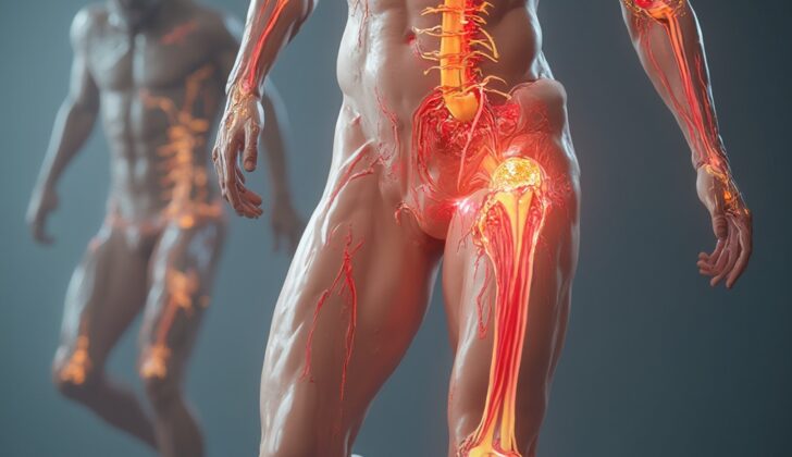
The femur, or thigh bone, has a complex structure. At the top end, the femur has a specially formed region, which includes the head, neck, and two protrusions called the greater and lesser trochanters. At the lower end, it transforms into wider structures known as the medial and lateral femoral condyles, separated by a gap. Between these two ends is the shaft. The first 5 cm below the lesser trochanter is a sub-region which tends to be challenging to manage when fractured due to the way in which the muscles pull on it.
The shaft of the femur is a smooth cylinder and areas of cortical thickness vary along its length, which can give an idea of femoral rotation during surgery. The femur is slightly curved forwards, with an average curve radius of 120 cm, and the curve increases as the radius shortens. The linea aspera, a ridge that runs along the posterior side of the femur, serves as an attachment site for muscles and acts as a structural reinforcement for the bone.
The femur is surrounded by several muscle groups. The muscles at the front are responsible for extending the knee and contain the femoral nerve. The rear muscles are for flexing the knee and contain the sciatic nerve, which is at risk of injury in femoral shaft fractures since it’s near to the shaft. The medial muscles are adductors, and the gluteal muscles located at the thigh’s side attach to the femur and cover certain nerve areas. When it comes to femoral shaft fractures, these muscles can change the alignment of the fracture pieces. In general, the upper segment is bent outwards and rotated by the iliopsoas and hip abductors. The lower segment is pulled upwards (shortened) by several other muscles.
The main blood supply to the femur comes from the femoral artery. This artery, which originates from the external iliac artery, splits into the superficial femoral artery (supplying blood to tissues below the knee) and the deep femoral artery (supplying the femoral shaft and surrounding tissues). It has many branches, notably the perforating arteries which encircle the femur. Additionally, nutrient arteries, which stem from the deep femoral or its branches, supply the inner two-thirds of the femur’s protective layer and bone marrow. These nutrient arteries connect with the metaphyseal-epiphyseal system. The outer third of the hard bone layer is supplied by the periosteal blood supply.
What Causes Femoral Shaft Fractures?
Femoral shaft fractures, or breaks in the main part of the thigh bone, can happen from a range of causes, both large or small in force. They often go hand-in-hand with other severe injuries. The usual culprits are car accidents, falls from a height, ordinary-level falls in people with weak bones, and gunshot wounds.
A study from Finland showed that 75% of these fractures were caused by strong impacts, with car accidents making up 87% of those cases (or 65% of all thigh bone fractures).
Other less frequent reasons for a broken thigh bone include unusual fractures from the use of a certain type of medication called bisphosphonates, fractures through an area of abnormal bone, fractures due to weakened bones from osteoporosis, and stress fractures caused by overexertion in athletes and military recruits.
Risk Factors and Frequency for Femoral Shaft Fractures
Femoral shaft fractures, or breaks in the thigh bone, happen around 10 to 21 times per 100,000 people every year worldwide. Only about 2% of these are open fractures, where the bone breaks through the skin. However, unusual thigh bone fractures as per the American Society for Bone and Mineral Research occur in anywhere from 3.5% to 16% of cases.
The chances of getting a thigh bone fracture are not the same throughout life. Men are more likely to get them between 15 to 35 years old, while women’s risk increases steadily from the age of 60. Men often get these fractures from car accidents or other intense events, while women often get them from falls from standing height. These injuries are therefore more common in younger people due to car accidents and in older people due to falls, thought to be linked to osteoporosis or weakened bones.
Type of fracture could also be related to the age at presentation, which has been increasing over time. For example, in the 1990s, the average age at presentation was 44, but it increased to 65 by 2000, possibly due to stricter traffic laws and safer cars.
- Femoral shaft fractures happen 10 to 21 times per 100,000 people each year worldwide.
- Only around 2% of these fractures are open fractures.
- Unusual thigh bone fractures make up about 3.5% to 16% of cases.
- Men often get these fractures between the ages of 15 and 35.
- Women’s risk starts to increase steadily from the age of 60.
- Common causes for men are car accidents or intense impacts.
- Common causes for women are falls from standing height.
- The average age at presentation in the 1990s was 44, but by 2000, it had increased to 65.
Signs and Symptoms of Femoral Shaft Fractures
Every patient who is injured, even if it’s from a fall on flat ground, should receive immediate medical attention to identify any serious health issues that could prevent proper treatment. People who have fractures in the shaft of their femur, or thigh bone, will typically experience pain, bruising, swelling, deformity, shortening of the leg, and instability around the thigh. In cases where a person has been injured in multiple areas, it’s crucial to perform a full examination because the other injuries can sometimes hide the symptoms of the fracture.
About 2% of all femoral shaft fractures are open fractures, which are very serious injuries. It’s important to thoroughly examine the patient to identify any open fractures. If they are present, the patient must be given antibiotics and a tetanus shot right away to prevent infection. Any visible debris should be removed immediately, and the wound should be covered with a sterile, saline-soaked bandage. The patient should then be taken to the operating room for more thorough cleaning and treatment of the wound. Open fractures can cause uncontrolled bleeding and increase the risk of infection. In fact, one study found that open fractures categorized as Gustillo-Anderson type III had an infection rate of 17.6%, while types I and II had a rate of only 2.3%.
Furthermore, open fractures don’t protect against compartment syndrome, which can occur due to blunt force and violent movement of the femur through the surrounding tissues. In a study looking at patients with thigh compartment syndrome, 48% were found to have femoral shaft fractures.
It’s imperative to document the patient’s neurovascular status. While it’s rare, vascular injuries can happen in up to 2% of femoral shaft fractures, especially those caused by gunshots or other penetrating trauma. Damage to the deep femoral artery (DFA) and its branches is most common, and it usually leads to significant bleeding rather than a decrease in blood flow because the thigh has plenty of collateral blood flow. Since the thigh can hold about 1.5 liters of blood, these vascular injuries can majorly impact a patient’s state of shock. In contrast, injuries to the superficial femoral artery (SFA) can cause reduced blood flow to the leg and foot.
If a vascular compromise is suspected, you should look for symptoms like no pulse, an expanding pulsating hematoma, a bruit, a thrill, hemorrhage, and acute ischemia. If any of these symptoms are present, the patient’s leg should be put in traction and an ankle-brachial index (ABI) test should be done. If the ABI is less than 0.9, the patient should receive a computed tomography (CT) angiogram and be evaluated by a vascular surgeon.
Testing for Femoral Shaft Fractures
In simple terms, fractures in the femur (thigh bone) can be easily identified due to the deformation and instability of the thigh. Yet, in some cases, the injuries can be less noticeable. To identify these types of injuries accurately, additional evaluation, and scans such as X-rays and CT scans, are required. This is especially important for patients who are unconscious or less responsive.
As a part of the emergency response procedure, doctors often perform X-rays of the chest and pelvis. Once the patient’s condition is stable, they will take multiple X-rays from different angles of the injured leg. They do this to understand the nature of the fracture more specifically. Also, these X-rays could potentially identify fractures in other areas like the hip joint, upper femur, upper tibia (shin bone), and kneecap. It might help doctors to spot a “floating knee” injury, where both the femur and tibia are fractured.
CT scans are not the first choice for femur injuries, but they are frequently used when a patient has multiple injuries. Such scans can uncover hidden injuries and help plan the surgery better. High-resolution imaging can detect fractures in the neck of the femur, which is the uppermost part connecting to the hip. If the CT scan is performed with a contrast agent, it can also help in finding any vascular (blood vessel) injuries that need to be treated quickly.
When it comes to categorizing femur fractures, doctors often use descriptive terms, defining the location, type, angle, comminution (breakage into pieces), rotation, and displacement of the fracture.
The AO/Orthopaedic Trauma Association classification is widely used due to its reliability and accuracy. It uses a coding system that creates 27 different fracture patterns. The most basic division of this system includes Simple, Wedge, and Complex types, each further subdivided into specific categories.
The less commonly used Winquist classification only describes the breakage of the bone’s outer layer and whether the bone can bear weight after being repaired by inserting a metal rod into the marrow of the bone. With modern techniques in bone repair and sophisticated designs of these metal rods, the bone is usually stabilized, and full weight-bearing is allowed after surgery.
Treatment Options for Femoral Shaft Fractures
Femoral shaft fractures can either be treated surgically or non-surgically. The method of treatment largely varies depending on where you are in the world. High-income countries tend to opt for surgical treatment via a technique known as intramedullary nailing, recognized as the gold standard. Other surgical options include plate osteosynthesis and external fixation. Meanwhile in lower-income countries, non-surgical methods such as splinting or casting are frequently used, either as a temporary measure or as the definite treatment.
Intramedullary Nailing
Intramedullary nailing is regarded as the best method for treating femoral shaft fractures and is commonly performed. It’s recommended that this procedure be carried out as soon as possible, ideally within 24 to 48 hours to lower the chances of potential complications like lung infections. This method is considered particularly beneficial for its ability to facilitate early weight-bearing, which is instrumental in helping to maintain muscle strength and mobility.
There are two types of procedures: Antegrade Nailing and Retrograde Nailing.
Antegrade Nailing
This constitutes the standard treatment and is underpinned by positive results when conducted within the first 24 hours. Some concerns exist regarding early treatment for patients with closed head injuries due to possible pulmonary complications. Despite this, studies indicate that complications don’t actually increase unless the patient previously had a head injury.
Retrograde Nailing
Retrograde nailing has recently gained popularity, with expected results being comparable to antegrade nailing. Main complaints related to this approach include knee pain.
Reaming
This technique is highly beneficial for intramedullary nailing as it provides both mechanical and biological benefits. It was previously thought to increase the rate of pulmonary complications, but this has been debunked.
Plate Osteosynthesis
This solution, typically used for patients with fractures extending to the proximal or distal femur, is usually the last resort due to complications caused.
External Fixation
This is usually used in severe cases, such as open fractures or vascular injuries, or when the patient is not stable enough for immediate definitive care. It also comes with a relatively high complication rate.
Traction
First responders quickly assess and apply one of several temporary traction devices to stabilize femoral injuries, restoring the gross length, alignment, and rotation. More effective and long-term traction systems like skin and skeletal traction are applied in hospital. Skeletal traction may be used as a definitive treatment in some cases.
Other Considerations
As the population ages, more people will seek hip and knee replacements or undergo surgeries for fractures. This will lead to an increase in femoral shaft fractures associated with prosthetics (splints/artificial limbs), presenting new treatment challenges that need specialized care. In these instances, open reduction internal fixation may be necessary, increasing stress on the patient’s body.
What else can Femoral Shaft Fractures be?
When a person hurts their thigh bone or ‘femoral shaft’, it’s usually quite obvious and easy to identify with scans or X-rays. However, it’s crucial for the doctor to also check for any related fractures in the pelvic area, upper or lower thigh bone, and shin bone. The treatment plan may change based on these additional fractures. If a patient is using a type of drug known as bisphosphonates, the doctors need to look closely for thickened outer layers of the bones on both sides and signs that a break might be about to happen.
There can be cases where patients might be confused, unresponsive, or unable to give clear information about their injury. This can sometimes result in a delay in diagnosis. Hence, careful examination by healthcare professionals is paramount in such cases.
What to expect with Femoral Shaft Fractures
Using a procedure known as reamed nailing, healing rates have been remarkably successful, with 100% retrograde and 99% antegrade, along with impressive functional results. Patients who receive this treatment early on have better outcomes with fewer complications and a lower risk of death. After the procedure, patients can gradually start to put weight on their leg as they can tolerate, which speeds up their rehabilitation and returns them to their normal mobility faster.
However, for older patients, recovery might take a bit longer and be more difficult, often due to several other health conditions they may have. Cases of patients with fractures in both femur bones are more likely to result in death, especially if they have other injuries or are physiologically unstable. In fact, the mortality rate jumps from 9.8% for those with isolated fractures in both femur bones to 31.6% when they also sustain other injuries.
Patients with a single femur fracture have a hospital mortality rate of 1.4%. However, if they don’t receive treatment within 48 hours, their chance of dying increases five times.
Possible Complications When Diagnosed with Femoral Shaft Fractures
During a surgical procedure, there can be a variety of complications. These may include damage to the nerves or blood vessels, unintentionally caused fractures, compartment syndrome which is a painful condition caused by pressure buildup from internal bleeding or swelling of tissues, thermal necrosis which is tissue death from excessive heat, and improper alignment of a broken bone. After surgery, complications can include fat emboli syndrome which is when fat tissue gets into your bloodstream and blocks blood flow, pulmonary embolism, when a blood clot gets lodged in the lungs, infections, osteomyelitis which is an infection in the bone, improper healing of the fracture, non-healing fractures, and pain in the hip and knee.
Excessive reaming, a procedure to clean out or enlarge a hole in the bone, can lead to high temperatures of up to 57 degrees C, causing tissue death and possibly delayed fracture healing. Studies on animals have shown that bone death can occur at temperatures of 47 degrees C if reaming takes too long.
There may be injuries to nerves, and these can be due to the patient’s position during surgery as well as unintentional damage from the surgeon. Pudendal nerve injuries, which affect the area between the legs, happen in around 15% of patients positioned with a perineal post. The sciatic nerve, running from the lower back to the feet, can be damaged during retrograde nailing with excessive pulling of the lower extremity or surgical instruments.
Vascular injury to the DFA (deep femoral artery) or SFA (superficial femoral artery) can happen after penetration with instruments or implants. Even when safe areas are used for inserting an implant, unusual anatomy can increase the patient’s risk of unintentionally caused injury.
Fractures occurring during surgery can happen, particularly of the greater trochanter (a part of the thighbone), the knee, and perforation of the anterior (front) cortex by the implant. Those with an unusually low curve in their femur are at increased risk for anterior perforation. After surgery, fractures are most common in areas where there’s more stress at the ends of an intramedullary nail or plate.
Improper rotation is one of the most severe complications of long bone fractures, occurring in up to 25% of cases. A rotation of more than 14 degrees from neutral can change the way a patient walks and the efficiency of their movement. Patients tend to cope better with internal rather than external misalignment. There are several ways to check for rotation, including cortical overlap and checking the rotation of the lesser trochanter with fluoroscopy. Computed tomography of both extremities is the most accurate way to check rotation.
Comminuted fractures, where the bone is broken into pieces, can pose a challenge in determining leg length that can lead to pelvic tilt causing hip and back pain. In a study of comminuted femur fractures, six patients had a leg length difference greater than 1.25 cm, with only 4 of these patients needing further surgery.
A nonunion fracture is when the fracture fails to heal, or when there’s a lack of signs of healing for six months. Non-surgical treatments may include taking supplements or vitamins D and calcium and external bone stimulation. The patient must be checked for possible infection as a cause of nonunion. Surgical treatment may include additional fixation with or without a bone graft, depending on why the fracture isn’t healing.
Recovery from Femoral Shaft Fractures
After surgery, patients may face local and systemic complications. Blood tests can detect issues such as anemia and kidney problems along with other metabolic disturbances. Doctors will look for problems like compartment syndrome, complications with the surgery wound, and issues with the nerves and blood vessels. Systemic complications include things like deep vein thrombosis, pulmonary embolism, and fat emboli syndrome.
Pretty soon after the surgery, patients will begin physical and occupational therapy in the hospital. The goal here is to help them regain movement and function for everyday tasks. This will continue even after they leave the hospital. Usually, patients are allowed to bear weight as they can tolerate and return to full or near-full normal function before the x-ray shows healing, this can be seen as callus formation on three out of the four sides of the bone structure.
Doctors recommend that patients avoid smoking and taking nonsteroidal anti-inflammatory drugs (such as ibuprofen), though it’s noted that the evidence against these drugs isn’t clear cut. When they can bear weight without help and use their right leg safely to brake, patients are generally allowed to drive again. Medical hardware used in the surgery generally stays in place until a specific need arises to remove it. Removal of part of an implant is considered acceptable in cases where visible screws are causing local irritation.
Preventing Femoral Shaft Fractures
It’s impossible to completely prevent events that cause fractures in the large bone in the thigh, known as femoral shaft fractures. However, society is working on reducing the number of these incidents. Every year, around 40,000 people die due to car accidents. These accidents are responsible for up to 87% of femoral shaft fractures. Governments are working towards making car travel safer for both passengers and pedestrians. They’re doing this by creating rules against impaired driving and requiring improved vehicle safety equipment and designs.
Doctors should also check their patients who are at risk of developing a brittle bone condition called osteoporosis. If they spot any signs, they should guide those patients towards the right medical treatment to prevent fractures.


