What is Galeazzi Fractures?
The forearm is a crucial part of the human body, helping us perform everyday activities. It’s specially designed to allow our hands to rotate inwards (pronation) and outwards (supination). Injuries to the forearm, such as fractures, can cause significant problems both immediately and in the long run, especially if not treated correctly.
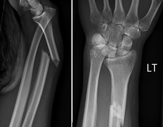
Sir Astley Cooper first talked about a specific kind of forearm fracture in 1822, but it didn’t get its name until 1934 when Riccardo Galeazzi explored the cause, frequency, and treatment of this injury. This fracture, now known as a Galeazzi fracture, happens in the middle to the end of the larger bone in the forearm, the radius. It’s often associated with a displacement of the joint where the radius and the smaller forearm bone, the ulna, meet at the wrist.
With advancements in imaging technology and fracture research, we can better understand, categorize, and plan treatment for Galeazzi fractures. Despite this, they’re still challenging to identify based on symptoms alone, and serious complications can arise if not properly treated.
What Causes Galeazzi Fractures?
Galeazzi fractures usually happen when someone falls onto an outstretched hand with their wrist extended and their forearm excessively rotated. The force from the break in the radius (a bone in the forearm) goes toward the joint between the radius and ulna, causing it to dislocate. This type of fracture tends to happen in two specific groups: young males who experience high-energy injuries like sports injuries, falls from high places, or car accidents, and older females who experience lower-energy injuries, like falling over from a standing position.
Risk Factors and Frequency for Galeazzi Fractures
Galeazzi fractures, a specific type of forearm fracture, make up about 7% of all adult forearm fractures. Out of every four radial shaft fractures, one is a true Galeazzi injury. Most forearm fractures occur in the distal area, the part furthest from the body, rather than in the midshaft area, which is closer to the body. Midshaft forearm fractures occur in 1 to 10 out of every 10,000 people each year.
These types of fractures are most common in children between the ages of nine and twelve. Playing sports like football and wrestling, suffering from osteoporosis, and being post-menopausal are all factors that can increase the risk of getting a midshaft forearm fracture. These risk factors result in the highest occurrence of this type of fracture in young males and elderly females.
- Galeazzi fractures make up about 7% of adult forearm fractures.
- Out of every four radial shaft fractures, one is a Galeazzi injury.
- Distal forearm fractures are more common than midshaft forearm fractures.
- Midshaft forearm fractures occur in 1 to 10 out of every 10,000 people each year.
- These fractures are most common in children between the ages of 9 and 12.
- Participating in sports like football and wrestling, suffering from osteoporosis, and being post-menopausal can increase your risk.
- The most common groups to get these fractures are young males and elderly females.
Signs and Symptoms of Galeazzi Fractures
People with forearm fractures typically feel pain at the point of injury. When examining these patients, doctors should first visually check the skin and surrounding tissue for any obvious damage. This can include deformities in the bone, cuts in the skin, bruised muscles, damaged tendons, and problems with blood flow or nerve function.
It’s really important to look out for any wounds that are directly above the fracture site, as this suggests an open fracture which requires immediate surgery. Doctors will carefully touch the area to further spot any deformities or sensitive areas. It’s also essential to check the stability of the joints above and below the fracture site for any additional injuries. For example, if a person falls on a hand spread out to break their fall, it can often mean a potential wrist injury, and special focus should be given to the stability of the Distal Radioulnar Joint (DRUJ).
Open wounds should not be tampered with. Severe crush injuries require a more thorough nerve and blood flow exam that might need to be repeated over time. This is done to look out for signs of acute compartment syndrome, a serious condition that can cut off blood flow to and from the arm.
Patients should also be asked about other symptoms like weakness, numbness, an abnormal prickling sensation, or pain that seems to be radiating from the injury site. Even though nerve damage isn’t very common, examining the areas catered to by the median and radial nerves is necessary to rule it out as much as possible. Ulnar nerve injury, another type of nerve damage, is even less common but should still be considered during the examination process.
Testing for Galeazzi Fractures
If you have a suspected broken forearm or a forearm bone slipping out of place, your doctor will likely order X-rays. Standard X-rays typically involve two views – one from the front (anteroposterior) and one from the side (lateral). Sometimes, an additional angled (oblique) view might be used to better understand the nature of the injury.
Extra X-rays of the wrist and elbow can also be taken if your doctor thinks you might have additional injuries in these areas.
If the X-rays show a fracture around the middle of your forearm (specifically, the radius bone, which is one of the two bones in the forearm), your doctor will check carefully for signs of injury to a joint called the distal radioulnar joint (DRUJ). Signs of this type of injury can include:
- Widening of the DRUJ seen on the front-on view
- Fracture of the ulna’s projection at the wrist (ulnar styloid)
- Displacement of the ulna backwards on the side view
- Shortening of the radius more than 5 mm (this would require a comparison with your uninjured arm)
Most of the time, these regular X-rays are enough for the initial evaluation, and no further imaging is needed. However, for planning surgery or if your bone hasn’t healed properly (non-union), a computed tomography (CT) scan could be performed. An MRI (magnetic resonance imaging) can also be used, which could reveal tears in the disc-shaped structure of cartilage in the wrist (TFCC) or damage in the membrane between the two forearm bones.
Treatment Options for Galeazzi Fractures
If someone is suspected or confirmed to have a Galeazzi fracture, which is a specific type of wrist fracture, they will need to be seen by a bone specialist, known as an orthopedist. While waiting to see this specialist, the patient’s arm should be kept in a special type of splint known as a sugar-tong splint. Younger patients are usually treated by using a method that doesn’t involve surgery, while adults often have to undergo a surgical procedure. This is because past cases have shown that non-surgical treatment in adults often results in poor recovery for over 90% of patients.
When treating a suspected fracture, the initial steps usually involve resting the injured area, applying ice to it, keeping it immobilized, and raising it. In most cases, the healthcare provider will try to physically restore the displaced bones of the wrist to their original locations in a process known as closed reduction.
Children often recover better from this type of injury compared to adults. Typically, children’s fractures are treated with closed reduction and splinting – a method of using a splint to hold the affected area in place. The splint is usually applied up to the elbow with the forearm being in a twisted up position. However, in cases where the injury is severe or complicated, the child may require surgery. This would involve opening up the affected area and fixing the bones internally, a method known as open reduction and internal fixation (ORIF).
Adults, on the other hand, tend not to recover as well with closed reduction and splinting. Numerous reports show that treating adults without surgery results in high rates of the bones failing to fuse and the bones moving out of place. Because of this, surgery is the preferred treatment method for adults. The broken bone in the wrist is first repaired firmly. If the wrist joint is stable after this, two wires are pinned to it. If the wrist joint is unstable, the disk in the wrist needs to be repaired, followed by pinning two wires. Sometimes, the bone at the tip of the forearm, called the ulnar styloid, may also be fractured and unstable. In such cases, the disk needs to be repaired first, followed by fixing the styloid using a special screw or wire. Patients who cannot have their joint relocated will require an open reduction procedure that involves removal of the tissue blocking the reduction and repair of the disk. After the surgery, these patients’ arms are placed in a splint or cast that covers up to the elbow.
The time it takes to recover from a fracture varies based on several factors such as how severe the injury is, how well the individual heals, and what the person intends to do with the affected arm. Rehabilitation normally starts six to eight weeks after surgery and is aimed at restoring full movement, fine motor skills, and painlessness in the arm. People who have physically demanding jobs or participate in sports may need more time in rehabilitation. Typically, people with less physically demanding activities can expect full recovery after 8 to 12 weeks. People with high-physical demand activities may need up to 12 to 16 weeks of rehab. Most patients usually have their surgical hardware kept in permanently, with only 10% needing to have it removed.
What else can Galeazzi Fractures be?
When a person has an emergency related to their arm, it could be due to a variety of issues. These may include:
- An elbow getting dislocated or broken
- The hand getting dislocated or one of the bones in the hand being fractured
- The wrist getting dislocated or one of the bones in the wrist getting fractured
All of these conditions require immediate medical attention and management to alleviate pain and prevent further complications.


