What is Genu Valgum?
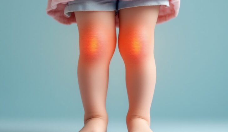
“Genu valgum”, also known as “knocked knees”, refers to a condition where the knees touch each other while standing straight, creating a distinct shape in the legs. Most people with knocked knees don’t experience any symptoms or problems with daily activities. Sometimes, however, it can be paired with flat feet and occasional inner foot and knee pain. This condition starts to develop in children around 2 years old, and it becomes most noticeable when they are between 3 and 4 years old. After that age, it usually lessens and settles into a stable, slightly outwards position by age 7. In teenage years, any changes in this condition are usually minimal.
To measure the severity of knocked knees, doctors use the “intermalleolar distance”, which is the space between the innermost points of the two ankles when a person is standing with their knees together. An intermalleolar distance of more than 8 centimeters (around 3 inches) is considered abnormal.
In rare cases, if the outward knee bending continues to increase, it could be associated with a turned-out walking pattern, a kneecap that slips to the side, and knees that rub together when the child walks.
What Causes Genu Valgum?
Bilateral Genu Valgum is a condition where both knees are bent inwards. This can happen due to several reasons, including:
Physiologic genu valgum, which means the condition is natural and usual for the growth stage.
Skeletal dysplasias, which refer to abnormalities in the growth and development of bones and cartilage.
Metabolic bone diseases, which are conditions that affect the strength of the bones and their ability to grow.
Lysosomal storage diseases, which are genetic disorders that cause a harmful buildup of substances in the body’s cells.
On the other hand, Unilateral Genu Valgum is when only one knee is bent inwards. This could happen for a few reasons, such as:
Post-traumatic, which means it is the result of an injury.
Tumors, which are abnormal growths of cells.
Infection, which is when harmful bacteria or viruses invade body tissues.
Risk Factors and Frequency for Genu Valgum
When examining the condition known as genu valgum, most patients are usually between the ages of 3 to 5. The primary area where this issue tends to occur is the lower part of the thigh bone, or femur. However, it can also happen in the shin bone, or tibia.
Signs and Symptoms of Genu Valgum
Kids between the ages of 3 and 5 commonly experience a condition known as knocked knees, properly called genu valgum. Parents usually notice this when their child’s knees touch but their ankles don’t. While this is often just a normal part of growth, it can sometimes be caused by more serious conditions.
Some of the conditions that may cause knocked knees include:
- Skeletal conditions like spondyloepiphyseal dysplasia and chondroectodermal dysplasia
- Metabolic bone diseases like rickets
- Lysosomal storage disease such as Morquio syndrome
When only one knee is affected (unilateral genu valgum), it’s often the result of trauma to the growth area of the bone. Doctors will use x-rays to look for signs of this trauma, like a narrower growth area or early closure of the growth plate.
The Cozen phenomenon is a specific type of trauma-induced knocked knee that can occur even with non-displaced fractures. The leading theory is the healing process of the fracture causes an overgrowth in the inner section of the bone. Other triggers for knocked knees can include radiation, infection, and tumors like osteochondromas, multiple hereditary exostoses, and fibrous dysplasia.
Testing for Genu Valgum
Looking at how someone walks and moves can tell us a lot about any physical issues they might be facing, like angular deformities (i.e., the legs aren’t straight). This is particularly important for children. There are different types of these deformities. For example, when the knee bends outward (valgus) during the stance part of the walk, it could be due to conditions such as renal osteodystrophy (a bone disease related to kidney problems) or an underdeveloped shinbone (fibula).
Another type of knee bend is called a valgus gait deviation which changes not just your side-to-side movement, but also your forward and backward movement. An example of this is increased femoral anteversion, where the thigh bone is tilted causing the leg to twist inwards creating an outward bend in the knee.
In children exhibiting normal growth patterns, x-rays aren’t necessary. However, if a child shows signs of excessive knee outward bend (genu valgum), has uneven limbs, falls into the height of the smallest 10% of their age group, or has a history of trauma or infection, then x-rays would be needed.
The x-rays begin with images of the legs under weight-bearing conditions with the kneecaps facing forward. These show any side-to-side deviations or twists in the lower legs which can then be measured. When we connect two dots: one at the center of the hip joint, and the other at the center of the ankle, this forms the “mechanical axis.” Normally, this line should pass through the center of the knee. If it deviates to the side, it can indicate conditions like genu varus (bowed legs) or genu valgum (knocked knees).
The tibiofemoral angle, formed between the shafts of the thigh and shin bones, changes as a child grows. At birth, the angle measures between 15 – 20 degrees, indicating bowed legs (varus angulation). By age 2, the legs straighten and then bends outwards (valgus angulation) between 10 – 15 degrees around ages 3 to 4. The legs then slowly move back towards straight, with a small 3 – 5 degree outward bend by the age of 7. This slight bend remains into adulthood and should not increase.
Determining whether the physical deformity originates in the thigh or shin bone is done by measuring two specific angles, namely, the mechanical lateral distal femoral angle and the medial proximal tibial angle. These two angles, formed in relation to the mechanical axis of the bones, should normally range between 85 and 90 degrees.
Treatment Options for Genu Valgum
For children under 6 years old displaying a natural, (or physiologic), “knock-kneed” condition also known as genu valgum, or where the angle of the lower leg to the thigh is less than 15 degrees, the usual recommendation is to simply observe the situation as it commonly rectifies itself over time. Wearing braces is not normally recommended for such cases. Moreover, if the “knock-kneed” condition is caused by trauma or injury, maximum distortion is often observed one year after the incident. However, monitoring the situation for 1 to 2 years is usually sufficient as most of these cases will resolve naturally without causing any impact on mobility. Furthermore, if metabolic disorders cause the distortion, the deformation can disappear upon treating the disorder, with medical management being the initial treatment approach.
In the case of larger angles, larger than 15 to 20 degrees, or if the midline of the body falls on the outer part of the upper shin bone (proximal tibia) in children less than ten years or those over ten years old, a corrective procedure known as hemiepiphysiodesis may be employed to guide proper growth. This procedure involves the use of implants such as screws, plates, or staples placed on the side of the bone, without disturbing the thin layer of tissue covering the bone (extraperiosteally). The Green-Anderson growth chart, which maps out the expected development of children’s bones, can help determine the timing of this procedure. Generally, younger patients experience quicker results. However, if the correction occurs too rapidly, the hardware may need to be removed to avoid overcorrecting to the opposite condition called varus (bow-leggedness). Due to a tendency for “bounce-back” growth following hemiepiphysiodesis, it could be beneficial to allow a slight degree of overcorrection before removing the implants.
Doctors closely watch these patients at 4 to 6-month intervals to ensure the effectiveness of the procedure.
For patients near or at the age of full skeletal maturity, another treatment option is a surgical procedure known as osteotomy. If the “knock-kneed” condition is primarily due to misalignment in the long bone of the thigh (distal femur), a type of osteotomy can be performed. One technique is to close a wedge on the inner side of the leg (medial closing wedge osteotomy) or create an opening on the outer part (lateral opening wedge osteotomy). An external fixator, a device used to stabilize the bones, may also be used if the patient requires lengthening the leg as well.
What else can Genu Valgum be?
Doctors need to tell the difference between normal knock-knees and knock-knees that are a result of a disease or disorder. This is particularly important if the knock-knees are not symmetrical, if they seem extreme, or if they occur in people who are older than the usual age range for this condition. It’s also crucial to look into this if the person is shorter than 90% of people their age, or if they have had a previous injury or infection.
What to expect with Genu Valgum
“Physiologic genu valgum” and “Cozen’s phenomenon” refer to conditions that cause an individual’s knees to bend inward. These conditions usually correct themselves on their own over time. If the genu valgum is associated with disorders affecting the metabolism of the bones, treating these disorders often brings improvement.
However, it is still unknown at what rate and frequency “pediatric genu valgum” – which happens in children – leads to adult degenerative joint disease, a condition where the cartilage around the joints breaks down over time.
Possible Complications When Diagnosed with Genu Valgum
If genu valgum, or knock-knees, is not properly diagnosed, certain complications can occur. These are typically related to the root cause of the condition.
When surgical intervention is involved, there can be further complications:
- Injuries to the physis, the growth plate in the bone, when implants are placed under the skin’s outer layer
- Over or under correction of the knees alignement
- Inadvertent damage to the nerves or blood vessels
- Infection
Preventing Genu Valgum
Most kids come to the clinic between the ages of 3 to 5 because their parents are worried about the appearance of their bent knees. It’s important for parents to know that this condition, known as genu valgum, commonly occurs in this age group and usually gets better on its own as the child grows older.


