What is March Fracture (Metatarsal Stress Fractures)?
March fractures, also known as metatarsal stress fractures, were originally identified back in 1855 when Prussian soldiers experienced foot pain and swelling after long marches. These are fractures in the metatarsal bones of the foot caused by repeated stress. Both individual health factors and external environmental factors can contribute to these fractures occurring.
To diagnose these fractures, doctors usually look at a patient’s medical history, perform a physical examination, and use imaging like X-rays. However, it’s worth noting that patients often show early signs of these fractures even before they can be seen on an X-ray. X-rays might not show the fracture for 2 to 4 weeks after symptoms start.
Typically, these fractures are treated conservatively, which means without surgery. However, they sometimes fail to heal, a condition known as nonunion. In these situations, surgery might be required to fix the bones.
What Causes March Fracture (Metatarsal Stress Fractures)?
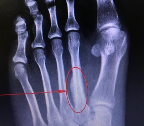
March fractures, also known as metatarsal stress fractures, are often seen in the second and third metatarsals located in the foot. These fractures are typically caused by overuse injuries. When there’s a sudden increase in physical activity in terms of intensity, duration, and frequency without enough rest, these fractures can occur.
Repetitive impact on the metatarsals, such as in weight-bearing exercises, can cause small cracks or microfractures. Over time, these small fractures can join together to create a stress fracture. This type of injury doesn’t come from one traumatic event, but rather develops over time due to continuous strain.
The most common place for metatarsal stress fractures to occur is around the neck of the second metatarsal. The second metatarsal is particularly vulnerable to injury due to its limited motion and its length compared to the other metatarsals in the foot. This is because it’s strongly attached by ligaments to the first and second cuneiform bones in the foot.
Risk Factors and Frequency for March Fracture (Metatarsal Stress Fractures)
Metatarsal stress fractures, or breaks in the foot bones, are fairly common, especially amongst athletes and military personnel. They can make up a quarter of all stress fractures and prompt one in five visits to sports medicine clinics. It’s estimated that almost half of all athletes will face a stress fracture at some point. Women, in particular, seem to experience these fractures more frequently than men. What’s noteworthy is that if someone has already had a stress fracture, they are typically more prone to having another one. In fact, 60% of those with a stress fracture have previously had another one.
- Metatarsal stress fractures are common, especially among athletes and military personnel.
- They account for 25% of all stress fractures and 20% of sports medicine clinic visits.
- About 40% of all athletes will experience a stress fracture in their careers.
- Women tend to have these fractures more frequently than men.
- If someone has had a stress fracture before, they’re more likely to have another one.
- Sixty percent of people with a stress fracture have had a previous one.
Signs and Symptoms of March Fracture (Metatarsal Stress Fractures)
During a medical interview, patients may express a gradual new onset of pain that briefly gets better with rest, but intensifies with physical activity. Doctors should ask about any recent changes in exercise routines, particularly any increase in intensity, duration, or frequency over the past 6 to 8 weeks. Other important factors include changes in terrain or footwear, as these can alter how force is distributed through the foot. The pain may vary in location but is commonly described as dull and aching, becoming worse during weight-bearing activities.
It’s necessary to take a comprehensive medical history, paying close attention to any risk factors. Intrinsic risk factors may involve age, endocrine disorders, body composition, bone mineral density, and past stress fractures. Older age and a low body mass index, particularly in women, are connected to higher rates of stress fractures. A previous stress fracture increases the risk of a future one due to the presence of related intrinsic factors or changes in bone structure as a result of past injuries. The questioning about extrinsic risk factors should involve more than just changes in activity levels. It should cover dietary habits, eating behavior, medication usage, and foot biomechanics. The female triad – characterized by irregular periods, low bone mass, and disordered eating – is a significant risk factor for stress fractures. Certain medications, such as bisphosphonates and glucocorticoids, can increase the risk of metatarsal fractures.
The physical examination should include a thorough biomechanical evaluation, involving palpation and pinpointing the location of the pain. If the fracture is near a joint, movement of that joint will likely worsen the pain. There may also be signs of a limping gait when the patient bears weight. The type of foot should also be examined, with a focus on a more supinated or less pronated foot, which carries a higher risk for second metatarsal stress fracture due to increased load across the forefoot.
Testing for March Fracture (Metatarsal Stress Fractures)
March fractures are typically diagnosed based on a person’s medical history and a physical examination, but diagnostic imaging is used to confirm the diagnosis. The initial step is usually plain radiographs, also known as X-rays. However, X-rays may not always immediately identify fractures in the metatarsal bones, often only showing fractures 2 to 4 weeks after the patient begins to experience pain. Sometimes, only subtle changes in the bone, such as a slight blurring, indicate a potential stress fracture.
If an expected fracture isn’t showing up on X-rays, other imaging techniques can be used. These techniques include bone scans, magnetic resonance imaging (MRI), and computed tomography (CT). Both bone scans and MRIs can detect fractures as early as 24 hours after the onset of pain. Bone scans are known to be sensitive to these fractures but might not be specific enough. However, MRI is considered the gold standard of diagnosis as it’s very accurate for detecting metatarsal stress fractures.
In addition to these scans, a CT scan can show features of the fracture but is not as sensitive as an MRI. Additionally, other tests may also be necessary to examine the patient’s individual risk factors. For example, doctors may measure vitamin D levels in patients who are suspected of having nutritional deficiencies.
Treatment Options for March Fracture (Metatarsal Stress Fractures)
In most cases, rest and pain relief are all that’s needed to heal a metatarsal stress fracture in the foot. Over-the-counter pain relievers like acetaminophen, along with icing the region, can help with pain and any swelling. The use of nonsteroidal anti-inflammatory drugs (known as NSAIDs) in helping these fractures heal is still a matter of debate.
Typically, there’s no need to immobilise the foot but a special stiff-soled boot might be worn for about one to two months. Patients are often allowed to put weight on the foot as long as it doesn’t cause pain. Low-impact physical activities like swimming, cycling or deep-water running can be added to exercise routines during the recovery phase. However, fractures occurring at the base of the foot’s fifth toe and the neck of the second toe are more likely not to heal properly. In cases where these fracture sites don’t show up on advanced imaging but are causing pain, a period of not bearing weight on the foot might be advised.
To promote healing and lower the risk of another injury, it’s important to address any factors that can be changed. This could involve taking supplements if there is a deficiency in vitamins D and calcium, or not getting enough calories. Slowing down the pace, duration, and frequency of training, as well as considering the use of supportive footwear or shock-absorbing insoles, can help to adjust and distribute the load more evenly during weight-bearing exercises.
What else can March Fracture (Metatarsal Stress Fractures) be?
When trying to diagnose a march fracture, which is a stress fracture in the foot typically caused by excessive walking or running, medical professionals need to rule out the following:
- Acute metatarsal or sesamoid fractures (breaks in foot bones)
- Injuries to nerves in the foot and leg, such as the common peroneal nerve, posterior tibial nerve, saphenous nerve, or sural nerve
- Conditions affecting the big toe joint, like hallux rigidus
- Specific types of fractures, such as a Jones fracture (a break in the outer part of the foot) or a proximal fifth metatarsal avulsion fracture (where a piece of bone is torn off)
- Stress fractures in the sesamoid bones (small bones in the foot)
Advanced medical imaging can sometimes make stress fractures appear similar to other medical conditions, like certain types of cancer that affect the bones (like osteosarcoma or Ewing sarcoma), metastasis (spread of cancer), chronic osteomyelitis (bone infection), or osteoid osteoma (a benign bone tumor). However, using MRI technology, doctors can accurately tell the difference between a stress fracture and a pathological one (due to disease) 93% to 98% of the time.
What to expect with March Fracture (Metatarsal Stress Fractures)
While most stress fractures recover through traditional care and rest, it’s important to effectively manage both internal and external factors. Doing so provides patients with the best opportunity to prevent slow healing, improper bone union, or recurring injuries.
Possible Complications When Diagnosed with March Fracture (Metatarsal Stress Fractures)
If someone is suffering from a stress fracture and there’s no sign of it getting better after 6 to 8 weeks, even with pain management and advanced imaging, it may be time to visit a foot and ankle surgeon. A common complication of march fractures (stress fractures caused by high activity levels) is nonunion, a scenario where the fracture fails to heal. Symptoms for this condition may include chronic pain, swelling, or instability, and it can affect anywhere from 20% to 67% of patients.
In cases like these, different surgical options may be offered including:
- Medullary curettage: a procedure to remove abnormal tissue from the bone marrow
- Autologous bone grafting: Using one’s own bone to repair the fracture
- Bridge plating: using metal plates to stabilize the fracture
- Intramedullary nailing: inserting a metal rod down the center of the bone to stabilize it
Do note that the healing process after surgery can be quite lengthy, sometimes taking months or even years. Other treatments might include bone stimulation, shockwave therapy, or ultrasound. However, injectable bone cement options have not shown desirable results on their own, and while noninvasive options such as pulsed ultrasound may sound appealing, they have not effectively sped up the healing process. Shockwave therapy, on the other hand, can be a promising noninvasive addition to other treatment methods because of its ability to increase bone turnover, stimulate osteoblasts (cells that form new bone), and increase new blood vessel formation.
Recovery from March Fracture (Metatarsal Stress Fractures)
People are advised not to go back to exercising until they have experienced no pain for five straight days during their normal daily tasks. As a March fracture – which is a type of stress fracture in the foot – heals, it’s important to gradually build up activity levels again. When the person can bear their full weight and exercise lightly without pain, they can slowly increase their workout intensity by 10% every week. This helps to ensure that another stress fracture doesn’t occur.
Physical therapy can also assist with reducing swelling, improving strength and balance, and carefully guiding the return to full activity. To prevent March fractures, a structured workout schedule that includes plenty of rest and a steady rise in intensity is recommended. Special footwear and insoles specifically designed for the activity can also help to protect the foot’s vulnerable areas from biomechanical overload, which means too much pressure or force.
Preventing March Fracture (Metatarsal Stress Fractures)
March fractures are small breaks in the foot bones that don’t move out of their regular positions. These kinds of fractures can happen in the metatarsals, which are the long bones in the feet. Several causes can contribute to these fractures such as excessive use, rigorous training, wearing the wrong shoes, foot deformities like flat-footedness and overpronation (walking on the insides of your feet), thinning of the bones, and practicing sports incorrectly or on improper surfaces. If these fractures are not treated, they can lead to a complete break in the bone.
Symptoms of a march fracture might include pain, redness, swelling, and bruising. Usually, the symptoms get worse when weight is placed on the foot. X-rays and MRI scans of the foot can assist in diagnosing the condition. Standard treatments often involve getting plenty of rest, restraining mobility, reducing athletic activities, getting advice on healthy eating, and sometimes having surgery to secure and mend the fracture.
Preventing march fractures is possible by following a structured exercise routine that allows for ideal recovery and gradually increases intensity. Shoes and insoles that are specific to your activity might also help prevent overloading the foot areas that are most susceptible to injury.