What is Pediatric Lateral Humeral Condyle Fractures?
Fractures in the outer part of the elbow, known as lateral humeral condyle fractures, are the second most common type of fracture in children’s elbows. These injuries are special because they occur within the joint but are prone to movement and inability to heal due to the pull of the forearm muscles near this area. If not treated properly, these fractures can lead to changes in the shape of the elbow.
Usually, these injuries happen when a child falls on an outstretched hand. Common symptoms include a swollen and painful elbow, limited movement, and tenderness. Sometimes, a bruise on the outer part of the elbow may also be visible. Medical professionals use various classification systems to manage these injuries, considering surgery if the fracture is displaced by 2 millimeters or more. If the fracture is displaced, there is a higher chance it will not heal. Risks following surgery include non-healing, deformation, restricted blood supply, and infection.
The humerus, or arm bone, is the longest bone in the upper arm. It joins with the shoulder blade at the shoulder joint and with the forearm bones at the elbow joint. The top part of the humerus includes the head, larger and smaller bumps, and two neck areas. The middle part of the bone has a spot for the deltoid muscle attachment and a groove to hold the radial nerve and artery. The bone gets wider closer to the elbow, making two ridges. The bottom part includes two angular bumps, which are sandwiching the condylar area that forms the elbow joint with the forearm bones.
The head of the humerus fits into a cavity in the shoulder blade. The neck of the humerus is where the joint capsule of the shoulder attaches. The greater bump is a noticeable feature on the outer side of the humerus, while the lesser bump is at the front. Between these two bumps is a groove that contains the tendon of the biceps muscle.
The deltoid muscle attaches to the humerus at the deltoid tuberosity. The radial groove positions the radial nerve and artery. The elbow joint is shaped by the outer bump called the lateral epicondyle, which also serves as an attachment point for the forearm muscles. On the opposite side, the medial epicondyle is where the muscles responsible for forearm flexing attach.
What Causes Pediatric Lateral Humeral Condyle Fractures?
Fractures of the lateral humeral condyle, a part of the arm bone, are usually connected with trauma, typically due to falls. These injuries are most commonly seen when someone falls onto an outstretched hand with a straight arm and turned-up forearm.
Risk Factors and Frequency for Pediatric Lateral Humeral Condyle Fractures
This type of injury is the second most frequent type of elbow fracture seen in children, making up 12% to 20% of all upper arm fractures in this group. Kids between 4 and 10 years old, especially those who are 6, are the most likely to suffer from this injury. Interestingly, the injury happens more frequently on the left side. Boys account for approximately 67.4% of these injuries. Other injuries that often happen at the same time include dislocations of the elbow on the same side (11.4%, pointing either to the back and side or the back and middle) and other upper arm fractures on the same side (such as at the tip of the elbow [2.8%], in the forearm [1.5%], or near the inner knobby end of the elbow [1.2%]).
Signs and Symptoms of Pediatric Lateral Humeral Condyle Fractures
Falling on an outstretched hand often leads to injuries of the elbow, such as swelling, pain, and difficulty in moving the arm. You might also spot a bruise on the outer part of the elbow, which could suggest a severe fracture. It’s important that the entire upper arm is checked carefully. You’ll often find that the elbow is tender and that movement is limited.
When first seen by a doctor, a detailed check of blood flow and nerve function in the arm is necessary. This involves verifying the strength of the pulse in the arm, evaluating sensations in the fingers, and checking all finger movements. This is done to rule out serious conditions like compartment syndrome – a painful condition that decreases blood flow to the body’s tissues and nerves. If the skin is cut or looks like it’s being pulled from inside (skin tenting), this could indicate more severe injuries that need to be immediately addressed with surgery.
Supracondylar fractures, which occur at the very top of the bone in your upper arm, can present with similar symptoms. But these fractures are usually linked with worse elbow deformity and problems with blood flow and nerve function than injuries to the outer part of the elbow.
Testing for Pediatric Lateral Humeral Condyle Fractures
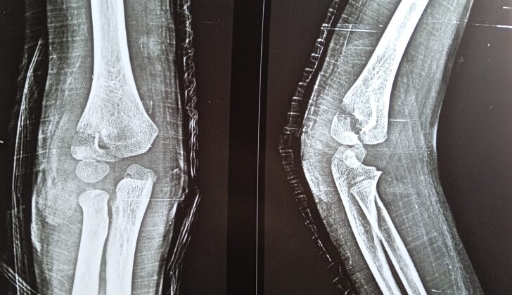
If you experience elbow pain, particularly after an injury, your doctor will probably start with an X-ray. The X-ray will include different views of your elbow from the front, side and an internal oblique view, which is a slanted view. This last view is particularly good at showing small breaks (less than 2 mm) that can happen when the bony bump on the outer edge of your elbow (the lateral condylar) breaks and moves slightly to the back.
An elbow arthrogram, which is a type of X-ray used in conjunction with a liquid dye injected into your joint, can also help confirm the exact problem and can be used during surgery to confirm the position of the bones. This technique is especially handy in cases where the broken pieces have shifted very slightly and can be treated with non-surgical methods.
Your doctor may also decide to use a computed tomography (CT) scan, which uses a series of X-rays to create a more detailed image. Conventional CT scans are a good tool for understanding how the fracture looks, but can’t give a great image of the cartilage, which is the softer material between your bones. Multidetector CT scans give even more detail than conventional CT and can help your doctor make treatment decisions.
Magnetic resonance imaging (MRI) can show the integrity of the cartilage. If the cartilage in the elbow is intact, the bone is generally still stable and less likely to shift later. But MRI is not commonly used because it can require child sedation and is more expensive. Ultrasonography, which uses sound waves to create images of the inside of the body, might be used instead to look for bone displacement in young children. Additionally, elbow arthroscopy, a procedure in which a small camera is inserted into the joint, can also help with diagnosis and reducing the fracture.
There have been many suggestions for classifying different types of elbow fractures in children. One, called the “Milch” classification, separates them into two categories based on whether the fracture extends to a particular part of the bone. But, this doesn’t always predict how the elbow will look during surgery and doesn’t give a lot of guidance on treatment. Another, by Jakob and Weiss, splits fractures into three types based on how much the bones shift and how intact the cartilage remains. A later one, by Song et al, describes five stages of fractures based on how they look, how much they shift, and how stable they are.
Treatment Options for Pediatric Lateral Humeral Condyle Fractures
For less severe fractures in children, where the displaced bone is less than 2 mm, a non-surgical treatment may be advised. This would involve immobilizing the arm with an above-elbow cast for at least 4 weeks, and maybe an additional 2 weeks depending on how the healing process is going. Doctors will typically confirm the healing process with x-rays both after the first week of treatment and then again on a regular basis to ensure the bone isn’t moving out of place.
However, if the bone displacement is larger than 2 mm, an open wound is present, or the healing process is slowing down, this would typically warrant surgical intervention. Evidence suggests that a delay in surgery of over 24 hours after the injury doesn’t affect the final results, though it might require a larger incision due to increased swelling. In such cases, doctors often prefer a minimally invasive approach using a technique known as percutaneous fixation – insertion of a pin or screw to stabilize the bone, which is commonly applied for certain types of fractures.
When using this pinning technique, using two pins instead of three can help improve the healing process by providing stability and reducing complications. The pins are typically removed 4 to 6 weeks after surgery, once x-rays and clinical assessment confirm the fracture has healed.
Several studies demonstrate the benefits of different practices in this area. For example, using ultrasound to help guide the placement of the pins may be more effective than other methods in avoiding open surgery. Also, while regular metallic pins and biodegradable pins yield similar results, biodegradable pins can offer additional benefits since they don’t need to be removed by a secondary procedure and they present fewer longer-term complications.
Also, alternative techniques like using cannulated screws that sit within the bone could be chosen as another option for fixation. While screws may need to be removed by additional surgery, they may help prevent skin infections that can occur with the use of pins.
However, if minimally invasive methods don’t work effectively, open reduction and internal fixation (also known as ORIF, a surgery to stabilize broken bones) might be required. Studies suggest that an anterolateral approach (accessing the fracture site from the front and side) yields better results than a posterolateral approach (from the back and side).
Interestingly, pinning remains an effective solution even in these more complex scenarios. Research has shown that smaller procedures that allow for pin insertion and larger surgeries that operate on the bone directly both yield satisfactory results. However, pinning can have advantages, such as minimal scarring and no need for a secondary surgery to remove the hardware used to stabilize the bone. Some recent studies even suggest that pinning techniques could be as effective and safe as larger surgeries in treating certain types of fractures while also avoiding visible scars.
What else can Pediatric Lateral Humeral Condyle Fractures be?
When checking for fractures in the outer part of a child’s elbow (known as lateral condylar fractures), doctors need to make sure they’re not dealing with a few other similar conditions. These include:
- Other injuries to the same arm
- Fractures above the elbow
- Elbow dislocations that are to the back and either to the side or in the middle of the arm
Each of these situations require a different type of treatment and can be identified with a detailed examination.
What to expect with Pediatric Lateral Humeral Condyle Fractures
The future outlook for children with lateral condyle fractures – fractures on the outer region of the elbow – is generally good when identified and treated quickly. According to studies, 91% of these fractures successfully heal. However, a late diagnosis can lead to future limitations in moving the joint and a condition called avascular necrosis, where bone tissue dies due to lack of blood supply.
The prognosis largely depends on accurately identifying the type of fracture and the suitability of the treatment method. The degree to which the bone is moved out of line – also known as fracture displacement – greatly affects the success of treatment. If the bone has moved less than 2mm, it is often recommended to treat it without surgery, as it usually results in fewer complications.
Possible Complications When Diagnosed with Pediatric Lateral Humeral Condyle Fractures
Fractures can lead to various complications, and the severity of the fracture often determines the complication rate. For example, Weiss type II fractures that require surgery have a lower complication rate (11%) compared to Weiss type III injuries (34%). A common complication in about 27% of cases is a prominent lateral condyle, but it majorly doesn’t affect the final recovery or health outcomes.
Unfortunately, if the broken bone pieces join in an incorrect position, a condition known as fracture malunion can occur. This can even lead to arthritis in the elbow joint if the articular surfaces are not properly aligned. Incorrect joining of fracture may also lead to deformities like cubitus varus (an inward angle of the forearm) or cubitus valgus (an outward angle of the forearm).
Lateral condyle fractures, a specific type of fracture around the elbow, can halt growth if the fracture affects the growth center of the bone. Additionally, abnormalities like fishtail deformities or avascular necrosis (bone death due to lack of blood supply) can occur in 14% and 1.7% of cases, respectively. These complications can particularly happen without proper communication between the different ossification centers (growth areas) in the bone. However, using techniques like lag screw osteosynthesis can help prevent these complications, especially when open reduction (re-aligning of the bones) is needed.
Nonunion, where the fractured bone doesn’t heal, is relatively more common with lateral condyle fractures. This complication ranges from 1% to 5%, based on different definitions. An effective way to treat nonunion is through surgical intervention, which has been found to lead to successful healing in many cases.
Elbow deformities, such as cubitus valgus (outward angle of the forearm), can be corrected by various surgical techniques. Recently, medial trapezoidal osteotomy and techniques using screws and combined autologous tricortical iliac bone graft reconstruction have shown effectiveness in treating these deformities, especially in pediatric lateral humeral condyle fracture nonunion cases.
Healing of a lateral condyle fracture often results in a wider end of the arm bone (distal humerus). Unfortunately, more than 20% of patients can develop cubitus varus, and more than 10% can develop a valgus deformity. Both these conditions can affect the physiological carrying angle (normal angle) of our elbow. The cause of these deformities is usually due to the limb healing at a different angle after the fracture.
However, a condition called pseudo-cubitus varus is reported to be more common than true cubitus varus. Moreover, with each 3.7 mm increase in the interepicondylar width (distance between the two bony prominences on either side of the elbow), the risk of developing true cubitus varus increases.
Tardy ulnar nerve palsy, a delayed onset condition causing weakness or paralysis of the muscles in the hand, is commonly a result of nonunion of lateral condyle fractures. This can cause cubitus valgus deformity (outward angled forearm), leading to ulnar nerve stretching and nerve damage symptoms. This is usually late-onset and may lead to a shrinking of the hand muscles.
Recovery from Pediatric Lateral Humeral Condyle Fractures
Kirschner wires, which are used in certain bone surgeries, are usually removed after a period of 4 to 6 weeks. Following their removal, it’s advisable to use a cast for an additional 2 to 4 weeks. The immobilization of the affected area in a long-arm cast that extends above the elbow continues until there’s evidence of healing both clinically and on X-rays. Once the cast is removed, exercises to regain the range of motion can start, but activities involving contact sports and heavy lifting should be avoided for 1 to 2 months.
A research study led by Bernthal et al found that, on average, patients had a motion arc of 64° at the time the cast was removed. Their data also indicated that patients who underwent surgery had a slower recovery, although there weren’t notable differences beyond the 18th week. Elements like older age, longer periods of immobilization, and more severe injuries were also found to cause slower recovery rates.
Preventing Pediatric Lateral Humeral Condyle Fractures
To help prevent children from suffering lateral condyle fractures, or broken elbows, we want to lessen the chance of falls and create safe environments for them. A few ways we can do this include:
- Watching over children while they play
- Picking softer play areas that have age-appropriate equipment
- Making the home safer, like by padding sharp corners and placing safety gates on stairs
- Making sure kids wear helmets, the right shoes and appropriate uniforms when playing sports
- Signing kids up for sports programs that are run by qualified coaches who prioritize safe practices
- Encouraging activities that help improve children’s balance and coordination
- Reminding parents and caregivers about the importance of taking steps to prevent injuries
- Addressing any health issues that could make children’s bones weaker
- Working with schools to introduce educational programs about injury prevention, safety rules and the importance of telling adults about any injuries or accidents
While these actions can reduce the risk of children suffering elbow fractures, it’s not possible to prevent all injuries. If a child does get hurt, it is important for doctors, families, and the injured child to talk openly with each other about what to expect during treatment and recovery. They should also discuss possible complications and understand that the severity of the injury could affect future complications, even after surgery. The role of physical therapy and rehabilitation exercises in helping the child regain full use of their elbow should also be stressed. It’s worth noting that recovering from an elbow fracture can take a child up to a year.


