What is Radius and Ulnar Shaft Fractures?
The hand is essential for us to interact with the world around us, allowing us to physically handle objects. The forearm, with its bones and muscles, is what allows the hand to function in various ways. The two bones in the forearm enable bending and straightening of the elbow and wrist through special types of joints.
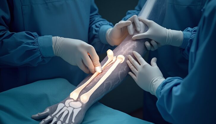
The two forearm bones, the radius and ulna, are positioned in a careful balance that lets us rotate our hand almost completely around. It’s the unique shape of the radius that allows this rotation, with the ulna remaining fixed in place. Any change to the forearm’s structure can significantly reduce the motion range, affecting both complex actions like a golf swing and simple tasks like turning a page in a book. The upper part of the ulna forms joints both with the lower part of the upper arm bone and with the upper part of the radius.
Several ligaments provide stability to the joint formed by the upper parts of the radius and ulna. These ligaments’ role changes with the motion of the forearm, and the stability they provide is critical for elbow stability. Similarly, the joint formed by the lower parts of the same bones is equally critical for wrist stability. There is a membrane joining the two forearm bones, which is vulnerable to injury during fractures. This text focuses on fractures affecting both the radius and ulna in adults, often called ‘both bone’ forearm fractures. While most common in children, these fractures also frequent in adults whose bones have stopped growing.
What Causes Radius and Ulnar Shaft Fractures?
Forearm fractures, which affect both bones, often happen in young people because of high-impact accidents or injuries. Common causes of these breaks in adults include car crashes, sporting accidents, and falls from a height.
Forearm fractures caused by less severe accidents, like falling over from standing, usually only happen in people who have weaker bones.
Risk Factors and Frequency for Radius and Ulnar Shaft Fractures
Fractures in the radius and ulna bones in the forearm are quite common, but there isn’t a lot of research defining their occurrence. These injuries tend to happen most frequently in two age groups: people under 40 and those over 60. Men and women have about the same risk of these fractures when they’re younger, but women over 60 are more likely to experience them. High school athletes and other active individuals also have an increased risk.
- Radius and ulna fractures in the forearm are common, but not well-studied.
- These fractures typically occur in people under 40 or over 60.
- Men and women have a similar risk of these fractures early in life.
- Women over 60 have a higher risk of these fractures.
- High school athletes and other active individuals are also at a higher risk.
Signs and Symptoms of Radius and Ulnar Shaft Fractures
Forearm pain after an intense injury is a common complaint. Just like with any physical injury, it’s essential to quickly rule out any life-threatening impacts. The starting point is often a step-by-step process to assess trauma, including a series of checks. Fractures involving both forearm bones usually cause a visible bending of the area and can sometimes break the skin. Therefore, a detailed examination of the skin around the fracture is critical.
If the skin is broken (known as an open fracture), it’s important to clean the area, administer antibiotics immediately, and give a tetanus vaccine if necessary. The best timing for cleaning and closing the wound is often disputed. A comprehensive examination of the nerves and blood vessels in the entire arm should also be carried out. This includes checking the function of specific nerves (AIN, PIN, and ulnar nerves) by having the person perform certain hand gestures and assessing the radial and ulnar arteries. It’s necessary to evaluate and record the blood flow to all fingers.
Although rare, associated injuries to the joint connecting the two bones of the forearm (the distal radioulnar joint) should always be checked by physical examination and X-ray. It’s also essential to check the front and back compartments of the forearm in the event of a severe forearm fracture. Data shows that a minor fraction of forearm fractures necessitated a surgical procedure to relieve tension or pressure (fasciotomy) due to suspected or confirmed compartment syndrome.
Compartment syndrome can occur even in open fractures. Several studies on acute compartment syndrome of the forearm have shown its presence even when there’s an open fracture.
Testing for Radius and Ulnar Shaft Fractures
If there’s suspicion of a forearm fracture, your doctor will begin evaluating it by capturing straight-on X-ray images of your forearm. These include views from the front (named anterior-posterior or AP) and side (called lateral views).
Sometimes, they might decide to capture images from an angle (referred as oblique views), or they could choose to also capture images of your wrist and elbow depending on your condition.
Typically, a CT scan isn’t necessary when dealing with forearm fractures. However, if your fracture seems complicated or there’s concern that it might have affected a joint (what doctors call “intraarticular involvement”), a CT scan can be beneficial.
Treatment Options for Radius and Ulnar Shaft Fractures
In children, non-surgical treatment of forearm fractures is common and usually results in good outcomes. However, in adults, non-surgical treatment of these fractures is rarely the best choice, and surgical treatment is often required. For example, fractures in the radial shaft (part of the forearm) generally need surgery to ensure proper healing and functionality.
On the other hand, if the ulnar shaft (another part of the forearm) has a minor fracture or is not displaced, it can usually be treated without surgery. This involves using a cast or functional brace, along with regular check-ups and examinations. But since most forearm fractures do require surgery, initial handling of the fracture should focus on getting the patient ready for the surgical procedure.
When fractures are significantly misplaced, sedative procedures can be used to effectively reposition the fracture and apply a splint. If the fracture is an open one, it is important to clean the wound immediately and start antibiotics as soon as possible. Splints, like the “sugar-tong” type, usually hold the forearm in a neutral rotation with the elbow flexed at a 90-degree angle. The surgical treatments available, typically involve either open reduction internal fixation (ORIF, where the bone is set in place openly and then fixated), or intramedullary nailing (inserting a nail into the center of the bone).
While intramedullary nailing can be quicker and cause less scarring, it can be difficult to stabilize the bone and restore its curvature using this method. Often, the ORIF method with plate and screw construct is considered the optimal treatment.
Some studies suggest a combination of plate fixation and intramedullary nailing is better, as it has good stability, fewer complications, and positive clinical results. The specific type of plate and screw used usually depends on the fracture pattern. Different methods are used based on the fracture patterns.
The aim of all these procedures is to restore the natural features and functionality of the bone. Bone grafting can also be used if there are segments missing, although its effect on fracture healing is still debated.
In case of open fractures, wound cleaning is performed at the time of surgery and prompt administration of antibiotics is critical. The extent of the soft tissue damage can determine the type of fixation method chosen. For severe open fractures with large soft tissue defects, temporary external fixation might be necessary before the final treatment and skin coverage.
What else can Radius and Ulnar Shaft Fractures be?
Determining if both bones in the forearm are fractured is generally a straightforward process with the right kind of imaging, like X-rays. By looking at the wrist and elbow, doctors can often rule out other associated injuries. It’s also important to keep an eye out for specific types of fractures known as Galeazzi and Monteggia fractures during the assessment.
The Galeazzi fracture involves a break of the lower third of the radius (one of the forearm bones) with an associated injury to the joint where the radius and ulna (the other forearm bone) meet further down the arm. On the other hand, a Monteggia fracture involves a break of the upper third of the ulna with the dislocation of the top part of the radius.
These types of fractures are most commonly seen in children. The treatment for these specific fractures differ from the usual treatment used for other forearm fractures.
What to expect with Radius and Ulnar Shaft Fractures
The outlook for patients with fractures of both bones in the forearm is usually excellent. Research has shown that a majority of these fractures fully heal within two months after surgical treatment. It’s recommended that patients start motion exercises for the wrist and elbow shortly after surgery, usually within a week.
Despite noticing slight reductions in grip strength and range of motion, the overall impact on the patient’s ability to perform daily tasks is minimal. In other words, patients’ rates of disability tend to be low following this type of fracture.
Possible Complications When Diagnosed with Radius and Ulnar Shaft Fractures
Acute compartment syndrome is a serious complication that can happen from both bone forearm fractures, whether treated with surgery or not. It means pressure builds in your muscles to dangerous levels. If this condition is suspected or confirmed, a critical surgical procedure called a fasciotomy needs to be done right away.
Surgery, especially due to these fractures, can also result in other complications like infection, bleeding, failure of the fractured bones to heal or heal in a misaligned way, injury to nerves and blood vessels. Ensuring the best medical state before surgery, using established surgical methods, and giving antibiotics during the perioperative period help in reducing these risks. Use of bone grafting, specifically in fractures with severe bone fragmentation, can reduce the risk of bones failing to unite.
The risk of the two forearm bones (radius and ulna) uniting abnormally is associated with a surgical approach that uses a single cut. Inadequate treatment or alignment of both bone forearm fractures can lead to a significant decline in mobility and function. Malunion, where the bones heal incorrectly, especially restricts the rotational movements of the forearm. The surgical hardware may cause discomfort and need to be removed. But taking out the plate and screws can increase the risk of re-fracture. Another important consideration is the condition of the fracture when first seen, early removal, lack of post-removal support, and the specifics related to the plate increase this risk. Injuries to joints in the forearm and elbow need to be suitably assessed and treated due to the unique complications related to such injuries.
Potential complications:
- Acute compartment syndrome
- Infection
- Bleeding
- Failure of fractured bones to heal
- Misalignments in healing
- Nerve and blood vessel injuries
- Unwanted union of the radius and ulna bones
- Loss of motion and function in the forearm
- Discomfort from surgical hardware
- Increased risk of re-fracture after hardware removal
- Complications due to injuries affecting joints in the forearm and elbow
Recovery from Radius and Ulnar Shaft Fractures
After surgery to fix fractures in the radius and ulna, or the long bones of the arm, the patient’s arm should be placed in a splint. This splint helps keep the elbow and forearm of the injured arm stable. However, the fingers and thumb are left free to move around, helping to avoid stiffness. About 5 to 7 days after the surgery, the patient can start exercises to move their elbow and forearm.
Patients need to have regular check-ups after the surgery until the soft tissues around the fracture have healed and the bones have united, which can be confirmed through X-rays. This recovery generally takes about 2 to 3 months. Once the doctor confirms that the bones have joined together correctly, the patient can then go back to doing most activities with the healed arm.
Preventing Radius and Ulnar Shaft Fractures
In situations where there’s a high chance of accidents, like driving a car, it’s always important to stay safe, for example, by wearing a seat belt. If an injury occurs, it’s important to immediately get to a trauma center for an assessment. It’s also crucial for people who sustain dual forearm fractures to understand what they might experience during healing and to recognize common complications. One such condition, called compartment syndrome, must be thoroughly explained.
Most patients will need their injured arm placed in a special protective covering called a splint and they will be asked to see an orthopedic surgeon for further follow-up. Instructions on how to take care of this splint will be given and the necessity of following up appointments will be emphasized. It’s also important to review and address any habits or behaviors that could make complications more likely at the first visit. This includes encouraging patients to quit smoking if that’s a relevant factor to them. Patients are advised not to put weight on the affected arm, and if there is a need for physical therapy, arrangements can be made during this time.


