What is Supracondylar Humerus Fractures?
Elbow fractures happen more often in children than in adults. This is mainly because kids tend to use their arms to break a fall, leading to a high number of elbow fractures. One specific type of fracture, known as a supracondylar humerus fracture, makes up almost 18% of all fractures in children and 60% of all elbow fractures. Dr. Gartland pointed out in 1959 that these elbow fractures can make even skilled trauma surgeons nervous due to their complexity. Despite improvements in how these fractures are evaluated and treated, they continue to present serious challenges for orthopedic surgeons.
Doctors frequently use a system known as the modified Gartland classification to identify the type of fracture and determine the best treatment. These injuries can be quite serious due to the risk of damage to the nerves, blood vessels, and potential for a condition known as compartment syndrome. If not set and held in place properly, the bone can heal incorrectly, leading to malformation and deformation. However, some patients who end up with such a malformation may still end up with satisfactory function in the long term.
What Causes Supracondylar Humerus Fractures?
The end of the upper arm bone, or humerus, is made up of inner and outer parts. These parts consist of the trochlea and the inner bump on one side, and the capitellum and the outer bump on the other side. The lower part of the humerus becomes narrower and thinner towards the middle of the arm.
The upper arm bone also gets narrower from front to back as the inner and outer parts merge at the top of the elbow pit. This creates a relatively weak spot in the area just above the bumps on the lower part of the upper arm bone.
Risk Factors and Frequency for Supracondylar Humerus Fractures
The area just above the elbow joint is where most elbow fractures happen in children, particularly between the ages of 5 and 7, with the average age being six. Although some studies suggest more boys or girls get these fractures, recent research shows no significant difference between the two. The most common type is the extension-type fracture, which often occurs when a child falls on an outstretched hand. The high flexibility of children’s elbows can also lead to such injuries.
- The area just above the elbow joint is the most fractured area in children.
- These fractures mostly occur in kids aged 5 to 7, with an average age of six.
- Boys and girls suffer from this type of fracture equally according to recent studies.
- Extension-type fractures are the most common, often caused by falls on outstretched hands or due to flexible elbows in children.
- These fractures are more common on weekends and during the summer season.
- Flexion-type fractures, more common in older children, frequently happen in the arm a child uses less often.
- About 1% of these fractures are open-with the bone exposed.
Signs and Symptoms of Supracondylar Humerus Fractures
If a person has fallen on an outstretched arm or a bent elbow, they might have a fracture. This type of injury is common, although in very rare instances (less than 0.5% of cases), the injury might not be accidental. Symptoms can include pain, swelling, or a noticeable change in the shape of the limb. Infants may not want to use the affected arm.
Physical examination is crucial to correctly diagnose and manage such injuries. The damaged arm should be inspected carefully for issues with the surrounding soft tissues and the blood vessels and nerves in the area. Any additional injuries should also be noted during the examination.
The injured arm might be swollen, show signs of bruising, and be deformed. Blood around the elbow could suggest an open fracture that needs to be assessed. A specific type of injury, known as “off-ended” Gartland III, typically shows an S-shaped deformity. Another sign to look out for is the “pucker sign” in the front of the elbow, which indicates a severe injury where the upper fragment of the broken bone has pierced through the muscle layer beneath the skin. This sign should raise concern for potential injury to the brachial artery or median nerve. It is also important to note, with such injuries, there’s a risk of developing compartment syndrome, a dangerous build-up of pressure in a muscle compartment.
Conducting a thorough assessment and documentation of the four major nerves in the area – the median, anterior interosseous, ulnar, and radial nerves is crucial. Both motor and sensory functions of each nerve should be documented. Significant nerve and blood vessel injuries can occur in these types of fractures – up to 49% in some cases. Vascular compromise happens in about 10-20% of serious elbow fractures. Traumatic neurapraxia – temporary loss of nerve function – is seen in around 11% of fractures, often involving the anterior interosseous nerve in extension-type fractures and the ulnar nerve in flexion-type injuries.
Brachial artery injury can occur in as many as 38% of serious fractures. Checking the blood supply to the limb is therefore crucial. This involves testing the radial and ulnar pulses, checking the temperature and colour of the same hand, and assessing the capillary refill time. It’s important to distinguish between a ‘pulseless pink, well-supplied hand’ and a ‘pulseless, pale, cold hand.’ Both scenarios require urgent medical attention, the latter indicating a dangerous shortage of blood supply, which is a surgical emergency.
Last but not least, neurological evaluation is paramount and should be repeated after every intervention. Although this can be challenging in children, certain tasks like playing rock, paper, scissors, and ok can be helpful.
- Rock (make a fist) – tests the median nerve
- Paper (extend the fingers) – tests the radial nerve
- Scissors (spread the fingers) – tests the ulnar nerve
- OK sign (thumb and index finger forming a circle) – tests the anterior interosseous nerve
Testing for Supracondylar Humerus Fractures
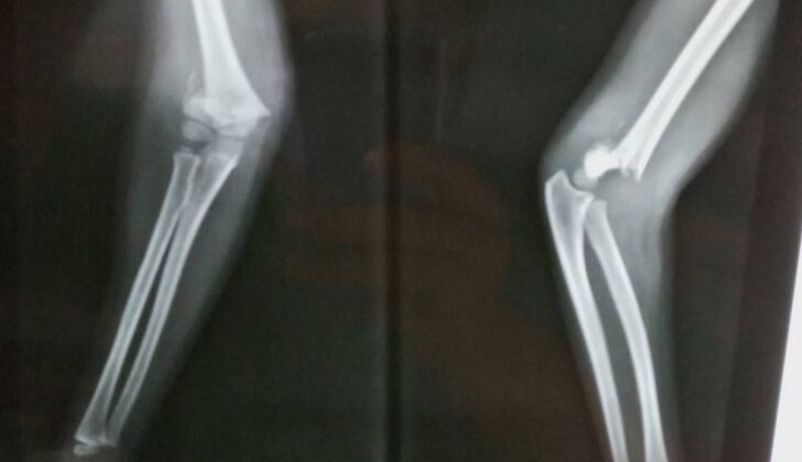
In order to properly diagnose an elbow injury, a basic x-ray of the elbow is required. Your doctor will need to look at the elbow from two different angles to confirm the type and extent of the injury. Understanding the growth process of the elbow, especially in children, is crucial to correctly reading these x-rays.
For the first type of x-ray (anteroposterior), the doctor will look for a fracture in the upper part of the elbow. Sometimes these fractures can be quite small and can be missed if the bone pieces haven’t shifted.
The doctor will also examine specific angles and lines in the elbow to assess the injury. Baumann’s Angle, for instance, is the angle formed by the arm bone and a line through the elbow joint. If this angle is increased, it can indicate a particular form of elbow deformity. Another check is the Radiocapitellar Line; a line drawn along the forearm should cross over a certain part of the elbow on all x-rays.
In the second type of x-ray (lateral), the doctor will look for the “posterior fat pad sign”. This is when a fracture causes internal bleeding that can push a small amount of fat up and away from a hollow in the elbow, indicating that a fracture has occurred. They’ll also examine the “anterior humeral line” which should cross through the middle of a particular elbow bone – if it doesn’t, this can indicate another form of fracture.
Although another type of imaging test (angiography) can be used, it’s usually not recommended if there’s a possibility that blood supply to the elbow has been compromised.
The severity of supracondylar fractures in children (fractures to the knob-like part of the upper arm bone) are labelled under Gartland’s classification. The fractures can be divided into extension-type or flexion-type injuries. Extension-type fractures are more common and can be sub-categorized depending on how displaced the bone is and whether there’s any rotational deformity or complete instability present at the fracture site.
Under the modified Gartland classification, the fractures are classified as follows: Type I is a non or slightly displaced fracture; Type IIA is a displaced fracture with the back part of the elbow intact; Type IIB is a displaced fracture with the back part of the elbow intact but a malrotation present; Type III is a completely displaced fracture with disruption of the back part of the bone skin; and Type IV is a completely displaced fracture with instability in many directions.
Treatment Options for Supracondylar Humerus Fractures
Advanced Trauma Life Support (ATLS) principles are used to assess all patients, and it’s vital to rule out non-accidental injuries in children.
Non-surgical treatment is suitable for certain types of fractures such as non-displaced Gartland I and minimally displaced Gartland IIA fractures. However, the treatment of Gartland II fractures can be controversial. These fractures can also be treated non-surgically if fracture alignment is desirable. But, there can be problems like misalignment of the fracture and malformation if there is a break in the medial column. There can also be risks of the medial column collapsing, leading to deformity. When there is too much swelling or posterior displacement, non-surgical treatment isn’t advised.
Non-surgical treatment generally involves immobilising the arm for three to four weeks using a collar and cuff or an above-elbow cast with the elbow flexed at 80 to 90 degrees. Excessive swelling might lead to compartment syndrome, a serious condition that involves increased pressure in a muscle compartment. Studies show that above-elbow casts can relieve pain more effectively than a collar and cuff. However, flexing the elbow too far should be avoided in patients with significant elbow swelling as it can also cause compartment syndrome. Historically, traction has been used for displaced fractures and can be used in settings where resources are limited.
Surgical treatment is required for displaced Gartland II and Gartland III fractures. Urgent surgical treatment is needed for patients with neurovascular issues, compartment syndrome and open fractures. The surgery generally involves reducing the fracture and percutaneously pinning with K-wires for closed injuries. Sometimes open reduction may be necessary. Surgeons should ensure they can handle any related injuries and have a vascular surgeon available for assistance during the surgery.
The closed reduction process involves traction and manipulation of the soft tissues to pull the fracture out to length. This is followed by adjusting the displacement and angulation of the distal fragment. Some rotations can be adjusted by pronation or supination, and also sagittal plane deformity can be corrected by hyperflexion. Sometimes the joystick technique might be used to reduce unstable fractures.
Percutaneous pinning technique involves using lateral and crossed pin constructs. Lateral pin configurations are often used to reduce the risk of nerve injury. Crossed pins increase risk of nerve injury, but they improve the rigidity of the construct more. It’s crucial to protect the nerve before inserting a medial pin.
If reduction can’t be achieved by closed methods, open reduction may be necessary, especially when the neurovascular structures are trapped in the fracture site preventing reduction. In such cases, vascular exploration is necessary. The patient must be monitored closely after the surgery.
What else can Supracondylar Humerus Fractures be?
In young children, a large part of the lower arm bone, or humerus, is made up of cartilage. As they grow, certain hardening areas known as ossification centers appear predictably around the elbow. However, in very young children, it may be difficult to make an exact diagnosis.
Conditions that could be considered when attempting to diagnose elbow issues in children include:
- Fractures of the radial head (the top of the radius bone in the forearm)
- Fractures of either the medial or lateral condyle of the humerus (the rounded ends of the bone)
- Transphyseal fractures of the humerus (breaks across the growth plate in the bone)
In cases of transphyseal fractures, it’s important to rule out non-accidental injury, or harm caused deliberately.
Another condition, commonly known as a “pulled elbow”, can usually be identified based on the child’s medical history, and X-rays or similar tests are typically not required in these cases.
What to expect with Supracondylar Humerus Fractures
The chances of full recovery after a bone injury greatly depend on three factors: the bone’s ability to heal and reshape itself, the patient’s personal health factors, and specifics about the injury itself.
The healing and reshaping ability of a bone is influenced by factors like the patient’s age, how much they still have to grow, how far the fracture is from the growth plate of the bone, the direction of the deformity, and how misaligned the bone fragments are. For example, the end of the upper arm bone provides 20% of the bone’s total growth. This portion of the bone won’t significantly reshape itself if it heals incorrectly. However, at the elbow, deformities in the front-to-back direction have a better chance of reshaping than those in the side-to-side direction. Younger patients who still have more growth left generally have a higher chance of their bone healing with the correct shape, particularly for injuries in the sagittal (front-to-back) plane.
Personal health factors also play a role. Children under the age of five tend to have better healing and reshaping abilities than those older than five. After age five, children’s upper arm bones have limited remaining growth. Additionally, obesity is often linked to more severe injuries and outcomes.
As for specifics about the injury itself, the severity of the injury, how well the bone is realigned and fixed, and the blood flow to the injured limb, are all crucial. Timely and appropriate treatment can significantly improve the long-term outlook of these injuries. Although worse outcomes may be associated with more severe injuries, correct realignment, stable fixation, and proper care of the surrounding soft tissues can enhance outcomes.
Minor fractures without displacement usually heal well and without complication. If not promptly identified, reduced, and stabilized, type II fractures with shattered fragments can result in a condition where the elbow angles away from the body. More severe type III and IV injuries are more likely to have associated nerve and blood vessel injuries, but with timely and appropriate treatment, the outcome can still be excellent.
Possible Complications When Diagnosed with Supracondylar Humerus Fractures
Complications from arm fractures can be broken down into several categories, each presenting its own potential issues:
- Neurological Injury: Traumatic neuropraxia can occur, which is a temporary nerve dysfunction due to impact. This damage can take different forms, such as Anterior Interosseous Nerve Palsy, Median Nerve Palsy, Radial Nerve Palsy, or Ulnar Nerve Palsy, depending on the specific location of the fracture.
- Vascular Injury: A common and serious injury is to major blood vessels, specifically the brachial artery, particularly in severe and complex fractures.
- Compartment Syndrome: As a result of severe vascular injury, pressure may build up in the muscle compartments of the forearm leading to what is called compartment syndrome.
- Volkmann Ischaemic Contracture: This is a rare but serious complication that can occur if compartment syndrome is not addressed in a timely manner and arterial damage is left untreated, causing muscle tissue to die off.
- Malunion and Deformity: If broken bones heal improperly, a host of deformities can ensue such as malunion, recurvatum, cubitus varus, or cubitus valgus, each presenting particular physical changes and complications. For example, Cubitus valgus can occur due to poorly aligned bone healing, which can lead to a late onset of ulnar nerve palsy – a condition that causes weakness, numbness, and pain in the hand and forearm.
- Stiffness: Limited arm mobility can frequently follow arm fractures, and is commonly associated with open surgery for repair, although range of motion often improves with time.
- Pin-site Infection: Infection at the site of surgical pins used in bone repair is a possible complication.
- Pin-site Migration: There’s also a risk that the surgical pins may move or shift within the bone.
Recovery from Supracondylar Humerus Fractures
After surgery, percutaneous pins, which are inserted through the skin, are typically taken out about 3 to 4 weeks later. As a result, kids can start to move around again. They may have some initial stiffness after the immobilization is removed, but this usually gets better quite quickly within the first month following the surgery. It’s worth noting that older kids may experience a slower return to their normal range of motion compared to younger ones. Studies have proven that physiotherapy does not necessarily enhance the recovery process, based on trials where patients were randomly assigned to receive or not receive this treatment.
Preventing Supracondylar Humerus Fractures
It’s important for children and their parents or caregivers to understand the severity and possible outcomes of their injuries. If the fracture is not displaced or only minimally displaced, there’s a good chance of a positive long-term outcome.
Displaced fractures, which are more severe injuries, may lead to a shape change of the affected area even after getting proper treatment. Sometimes, nerve injuries occur at the time of injury, but they often heal on their own without intervention. However, if this does not happen, further examination is necessary. Nerve injury can also happen during surgery, particularly when using a technique called percutaneous pinning.
Sometimes, a blood vessel injury can happen during the initial injury or during surgery. There are also other serious complications, like compartment syndrome, that should be brought to light. This requires a watchful eye. Starting the right treatment early can improve overall results, even for severe cases.


