What is Traumatic Lumbar Spondylolisthesis (Lower Spine Injury)?
Traumatic lumbar spondylolisthesis, also known as traumatic lumbar locked facet syndrome, is a medical term that describes an unusual injury where a bone in the lower back (ranging from L1 – L5) slips forward over the one below it. This tends to happen following a severe injury or trauma, causing significant changes at the joints between vertebrae – the small bones that make up the spine. Normally, lumbar spondylolisthesis is seen in people with age-related conditions.
Most often, injuries involving the traumatic shifting of the lower spine are found in the mid-to-lower back or the joint connecting the spine to the sacral region. In this article, we will explore the anatomy of the lower spine, focusing on the joints between vertebrae and how injuries can happen. We’ll also discuss the signs and symptoms, as well as what treatment options are available for this kind of injury.
What Causes Traumatic Lumbar Spondylolisthesis (Lower Spine Injury)?
The lumbar vertebrae, which are part of our spine, are made up of several parts including the vertebral body, two pedicles, two transverses processes, articular processes, lamina, and spinous process. This complicated structure can be broken down into three main sections.
The anterior column is essentially the vertebral body – a large block of bone with flat top and bottom surfaces, which supports about 80% of our body weight. The middle column is made of the pedicles. They’re connected to the posterior upper area of the vertebral body and act like a bridge, carrying tension and bending forces from the back to the front of our spine and supporting its stability.
The posterior column includes the transverse, spinous, and lamina processes, and the posterior articular processes. The transverse and spinous processes stick out laterally (to the sides) and medially (towards the middle) respectively, and are the sites where our back muscles attach. The laminas are a kind of protective bone layer that covers the neural parts of the vertebrae, extending from the spinous process to the posterior articular processes where they form the pars interarticularis. This separates the posterior articular processes into superior and inferior sections.
The vertebrae are connected to each other by an anterior intervertebral joint formed by an intervertebral disc and the posterior zygapophyseal joints. Here, the lumbar zygapophyseal joints are created by the combined superior and inferior articular processes of neighbouring vertebrae.
The arrangement of the zygapophyseal in the lumbar section of the spine is important to understand the movement related to spondylolisthesis, a spinal condition. Anatomically, the inside parts of the lumbar facets are arranged sideways (encouraging side-bending), while the outside facets resist rotation and side-bending. This arrangement shows a “C” or “J” shape from the side view and a straight line from the back view. Basically, the arrangement of the lumbar facet joints impacts their ability to resist forward slippage.
Various ligaments help to stabilize the lumbar spine by limiting its movement. The ligament flavum, supra-spinous, infra-spinous, and posterior longitudinal ligaments limit bending forward. Extension is limited by the anterior longitudinal ligament, while side bending is limited by the inter-transverse ligaments. The Ilio-lumbar ligaments provide additional resistance against forward movement of the L5 S1 section of the spine.
Muscles around the spine also contribute to its stabilization. These include the erector spinae, psoas major, and quadratus lomborum.
In light of this complex anatomy, it’s clear that traumatic spondylolisthesis involves a strong force that disrupts the muscle-ligament laying within the back.
Risk Factors and Frequency for Traumatic Lumbar Spondylolisthesis (Lower Spine Injury)
Traumatic lumbar spondylolisthesis is caused by intense accidents, such as car crashes or workplace accidents involving heavy objects making contact with the lower spine. This condition mostly affects males who are between the ages of 35 and 55.
Testing for Traumatic Lumbar Spondylolisthesis (Lower Spine Injury)
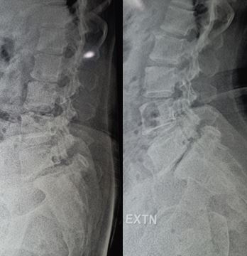
When the joint that connects two vertebrae (facet joint) gets disrupted due to injury, its lower part moves forward to sit on top of its upper part. This condition is known as “perched facet”. If the injury force is intense, the lower part moves even more forward, leading to a “locked facet” situation. These disturbances cause misalignment of the vertebrae, narrowing the spinal canal and potentially causing damage to the nerve roots or spinal cord.
These changes to the spinal structure can be easily seen on CT scans with 3D reconstruction or on MRI scans. MRI is particularly useful to visualize any trauma to the spinal cord or nerve roots as it can also show injuries to the muscles around the spine. A condition known as edema can occur due to strain or stretch injury to the muscles, and it is indicated by a brighter signal on the MRI images. Key features that doctors look for in these images include the loss of alignment at the facet joints and an increased distance between adjacent spine bones.
Doctors also need to assess the spinal discs irrespective of the vertebral alignment because serious disc injuries requiring surgery can be present even in the absence of any forward displacement of the vertebrae.
Spondylolisthesis, a condition where a vertebra slides forward over the one beneath it, is commonly graded using the Meyerding classification. This system divides the degree of forward movement of the vertebrae into five grades. Grades are as follows:
- Grade I: 0% to 25% forward movement (or low grade)
- Grade II: 26% to 50% forward movement
- Grade III: 51% to 75% forward movement
- Grade IV: 76% to 100% forward movement
- Grade V: more than 100% forward movement (also known as spondylosis)
Treatment Options for Traumatic Lumbar Spondylolisthesis (Lower Spine Injury)
The aim of the surgical procedure is to relieve pressure on the spinal canal and restore the stability of the spine. To achieve this, a procedure called laminectomy is performed, which involves removing part of the vertebra to create more space. If the vertebrae have slipped out of place, a technique involving manual traction and the removal of small joints in the spine called facetectomies may be required. This surgery helps correct the alignment of the vertebrae.
To further stabilize the spine, a procedure called posterior lumbar interbody fusion can be performed. This surgery involves using either open surgery or less invasive techniques to join the vertebrae together. After the operation, image testing usually shows satisfactory results, demonstrating that the pressure on the spine has been relieved and the spine’s stability has been restored.
What else can Traumatic Lumbar Spondylolisthesis (Lower Spine Injury) be?
If you’re feeling pain in your lower back, there can be several potential causes:
- Acute bone injuries in the lower back
- Disc injuries in the lower back
- A condition called facet syndrome that affects the joints in your lower back
- A sprain in the lower back
- Spondylolisthesis, which is when a bone in your back slides forward over the bone below it
- Spondylosis, which is a type of arthritis that affects your spine
- Myofascial pain, especially in athletes, which affects the tissue that surrounds your muscles
- Injury to the sacroiliac joint, which connects your spine to your pelvis
That might sound scary, but the right doctor can diagnose these issues and help you find relief.


