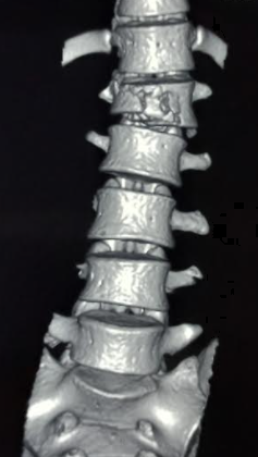
What is Vertebral Fracture?
The vertebrae are bones in your back that not only guard the spinal cord but also help in supporting your body weight and movements of the limbs. There are 33 of these bones in the spine. They are made in such a way that the soft, spongy bone is at the center and is enveloped by the solid, compact bone. The vertebrae also have structures called endplates that help in providing nutrients and help the disks attach to them. Essentially, these bones have what’s known as the body and a back arch. They also have two offshoots leading to a thin plate joining at the back, forming the spinous process. Furthermore, these bones come together to form a small opening which is crucial to the spine. There are also ligaments running down your spine, aiding in the movement and providing stability.
Each bone in the spine is connected to a disk on the top and bottom, which helps distribute body weight evenly. Other crucial parts of these bones include facet joints and processes that determine the spine’s movement. Understanding this complex structure is vital, especially when diagnosing and treating a potential vertebral fracture.
Vertebral fractures usually happen due to excessive weight loading, rotation or dislocation of the spine. This can occur due to accidents, bone-weakening conditions like osteoporosis, infection, or other bone diseases. For treatment, different systems have been designed to classify the severity and type of fracture. Usually, these systems consider factors like spinal stability, neurological damage, location, and extent of injury to the bones and linked ligament complexes. Despite the existence of many such systems, only a few have successfully achieved consistent reliability among medical professionals.
What Causes Vertebral Fracture?
Osteoporosis, a condition that weakens the bones, is the number one cause of fractures in the spine. However, there are also various other conditions and factors that can make the bones weaker and lead to these fractures. These include injuries, cancer, chemotherapy, infections, using steroids for a long time, overactive thyroid, and radiation therapy. Factors affecting bone density can be related to smoking or alcohol misuse, low levels of estrogen, eating disorders like anorexia, kidney disease, and some medications, including drugs that reduce stomach acid. Women, people over 50, people with a history of spinal fractures, smokers, people deficient in vitamin D, and long-term steroid users are at a higher risk.
Injuries are the second most common cause of spinal fracture, and the top cause of spinal cord injury is car accidents. Spinal cord injuries can occur anywhere from the uppermost part of the neck to the lower part of the back. Injuries below a certain point in the spine are referred to as cauda equina injuries. The National Spinal Cord Injury Statistic Center also includes falls and gunshot wounds as leading causes of spinal cord injury. Spine infections, like osteomyelitis, lead to hospital admissions in about 4.8 out of 100,000 patients.
Risk Factors and Frequency for Vertebral Fracture
Osteoporosis is a prevalent condition that affects over 200 million people worldwide. Up to 30% of women are affected by osteoporosis. Each year, more than 1.5 million Americans experience vertebral compression fractures, a common complication of osteoporosis. Women are almost twice as likely to have these fractures compared to men.
If a doctor diagnoses one vertebral compression fracture, your chances of having another fracture increases fivefold. If you’ve already had two fractures, your risk becomes twelve times higher. The consequences of these fractures can be serious and include both acute and chronic pain, a dip in the quality of life, loss of confidence, social isolation, and a higher risk of falls and subsequent fractures. These complications can also result in a doubled mortality rate compared to people without these issues.
The most common sites for compression fractures due to osteoporosis are between the T11 and L2 vertebrae located at the junction of the thoracic and lumbar spine. Moreover, on average, 160,000 traumatic spine fractures occur every year, with half of them affecting the same thoracolumbar junction. These types of fractures are more common in men, typically around the age of 30.

Signs and Symptoms of Vertebral Fracture
Vertebral compression fractures, often associated with osteoporosis, should be considered in both men and women with specific symptoms. Alarmingly, studies show that one in four women with these fractures goes undiagnosed. The main symptom is painful, either appearing suddenly or developing over time. This type of pain is often linked to activities that involve impact and puts pressure on the spine, such as standing or walking. The pain tends to lessen when lying down and increase when the affected area is touched.
Other symptoms and complications might also develop from vertebral compression fractures, including:
- Physical changes like back hump (kyphotic) or lumbar deformity
- Decreased ability to perform day-to-day tasks
- Constipation
- Reduced breathing capacity
- Deep vein thrombosis (clotting), often caused by prolonged lack of movement
- Social problems
Physical examination might reveal a loss of height, muscle loss, and change in the spine’s natural curve. Vertebral column fractures typically do not lead to symptoms like myelopathic signs (nerve symptoms) or radiating pain unless they are severe or cause restriction in the spinal canal.
Emergency care patients with possible spine injuries often undergo a CT scan of the spine after their breathing and heart rates are stabilized. Doctors also perform a comprehensive neurological evaluation, including checking the rectal tone, to detect possible spinal cord injuries. The evaluation is guided by clinical guidelines. In patients who are intubated and unable to participate in the exam, doctors assess reflexes and responses to painful stimuli.
It’s important to note that back pain is the most common symptom reported by patients with vertebral erosion due to infection or bone disease. Upon examination, doctors might palpate (feel) the spine to identify areas of concern which may need further imaging studies.
Testing for Vertebral Fracture
If elderly individuals with osteoporosis, or those at risk of osteoporosis, suffer from a fall or minor trauma and start complaining about acute back pain, a CT scan should be performed. This scan can check for spinal compression fractures, which are common in people with osteoporosis. Osteoporosis itself can also be diagnosed with a spinal bone fracture or through a DEXA scan showing a score of less than -2.5.
In the medical world, imaging, such as x-rays and CT scans, is considered the best method for evaluating spinal fractures. Specifically, a CT scan is the best way to examine spinal fractures as it enables the clinician to examine the spine from different angles and accurately determine the severity of the condition. A CT scan can also guide whether a DEXA scan is needed. An MRI can provide an image that shows how bones and soft tissues are interacting and how new or old the injury is.
For osteoporosis diagnosis, a Dual-energy X-ray absorptiometry (DEXA scan) is usually performed. The DEXA scan measures bone density, and a score less than -2.5 is typically indicative of osteoporosis. However, this scan isn’t useful in diagnosing compression fractures.
Biomarkers can also help diagnose osteoporosis but they cannot confirm compression fractures.
Various systems have been developed to label and grade spinal fractures. Studies have provided detailed classifications for the different types of land level, low impact induced spine fractures that could occur. These scales and classifications can also help in determining the severity of the injury and the appropriate treatment methods.
In cases where spinal fractures extend to the vertebral foramen, a specific CT angiogram might be needed to check for potential vertebral artery injury.
Patients who are suspected of having a spinal infection due to factors such as drug use, compromised immune systems, or diabetes should also be assessed. Medical professionals can use CT scans and MRI imaging to effectively evaluate for signs of osteomyelitis (bone infection) and discitis (disc inflammation) that could lead to spinal instability.
Treatment Options for Vertebral Fracture
Conservative treatment of acute osteoporotic compression fractures aims to ease pain and improve function. Typical strategies include painkillers such as acetaminophen, ibuprofen and opioids, and physical therapies like physiotherapy and rehabilitation programs. Bed rest and use of braces can also help with patient comfort. Wearing a semi-rigid thoracolumbar orthosis, a type of brace, has also been found to assist with walking in a small study. Ideally, successful treatment will address the original disease that caused the fracture in the spine.
When pain persists despite these strategies, surgical intervention may be required. A 2018 Cochrane review couldn’t strongly support the use of a procedure known as vertebroplasty for treating these fractures. However, long-term studies indicated that mortality rates are significantly higher for patients who only receive conservative treatment compared to those who have vertebroplasty or another procedure called balloon kyphoplasty.
In a vertebroplasty, doctors inject cement into the fractured vertebra via a needle under guided imaging, which then hardens and stabilizes the fracture. The procedure usually lasts one to two hours and patients can go home thereafter. Balloon kyphoplasty is similar, except a balloon is used to expand the vertebra before injecting the cement.
Management of spinal fractures aims to maintain spinal stability and preserve the function of the nervous system. Patients with spinal cord injuries (SCI) often need intensive care, including measures such as central and arterial lines for administering medications and possibly assistance with breathing. Trauma can cause hypotension, which can be counteracted with medications like norepinephrine or dopamine.
The objective of treating vertebral fractures is to attain clinical stability, assessed mechanically and neurologically. This involves ensuring the spine can withstand normal loads without causing further nervous system injury and preventing deformity and pain. Treatment will depend on the type of fracture, which might require surgery or conservative management.
Patients with infections of the bone (osteomyelitis) and spinal disc (discitis) typically receive intravenous antibiotics for 6 to 8 weeks, after obtaining a microbial diagnosis through biopsy. These conditions often don’t require surgery. Biopsy can also be used to diagnose metastatic disease, if it’s suspected.
What else can Vertebral Fracture be?
If an elderly individual is experiencing sudden back pain, it’s usually a good idea to get some sort of scan to check for fractures in the spinal column. If the scans do reveal fractures but there hasn’t been any kind of trauma or severe injury, then the underlying problem needs to be figured out. Some of the common causes can be osteoporosis, cancer that has spread to the bones, and other diseases that directly affect the bones. People who have been involved in serious accidents should also get scanned to see if there are any hidden injuries.
What to expect with Vertebral Fracture
Osteoporotic compression fractures, which occur when bones become too weak, can cause more fractures if left untreated. Treating this condition is crucial to prevent further damage to the spine. Two common treatments known as vertebroplasty and balloon kyphoplasty have been found to effectively reduce pain, decrease mortality and morbidity rates, and enhance the quality of life.
On the other hand, traumatic compression fractures – caused by a strong force such as a fall or heavy impact, often heal naturally and generally don’t need surgery. However, if the fracture is unstable, surgical stabilization becomes necessary. Additionally, early decompression can significantly improve outcomes for those suffering from incomplete spinal cord injuries, which are spinal cord injuries where some degree of sensation or motor function remains below the injury site.
Possible Complications When Diagnosed with Vertebral Fracture
Taking medicines for a lengthy period of time, like NSAIDs (non-steroidal anti-inflammatory drugs), due to conservative treatment methods can sometimes cause stomach ulcers and internal bleeding in the digestive system. Using opioids can also lead to changes in a person’s mental status and can potentially lead to addiction. If vertebral fractures are not treated, they may cause significant spinal deformities and continued degradation of the spine.
Pain can restrict an individual’s ability to move and can cause constipation, deep vein thrombosis (blood clot), pneumonia, weakening of physical capabilities, falls, and more fractures. Kyphoplasty, a procedure to fix fractured vertebrae, can also have complications such as cement leakage, spread of cement beyond the disc space, and cement blockages in blood vessels.
Surgery also has its own potential risks including but not limited to:
- Hardware failure
- Infections at the site of surgery
- Significant blood loss
- Neurological deficiencies
Preventing Vertebral Fracture
People who are dealing with fractures in their spine due to osteoporosis should focus on preventing any further fractures and treating their underlying health condition. This can help improve their overall wellbeing. Activities that can help strengthen the bones like weight-bearing exercises and muscle-strengthening routines are important. Besides this, adopting a healthier lifestyle can aid their recovery. This includes eating a balanced and nutritious diet, quitting smoking, and limiting the intake of alcohol.
Those who have osteoporosis might need to take certain medications like bisphosphonates or other drugs that tackle osteoporosis. This is done so as to lower the chances of experiencing more fractures. Patients who have had surgery or used a brace due to a traumatic injury need to regularly visit their doctor for check-ups in an outpatient setting. This way, the doctor can keep a close eye on their recovery progress.


