What is Acute Pneumothorax Evaluation and Treatment?
Pneumothorax is a condition where air or gas gathers in the space between the lungs and chest cavity. This can affect breathing and oxygen levels in the body, and its effects can range from being symptom-less to life-threatening.
This condition can be split into three main types based on how it is caused:
1. Traumatic Pneumothorax – This type is mostly due to chest injuries that may come from blunt force or piercing trauma. This is the most common cause of pneumothorax.
2. Iatrogenic Pneumothorax – This type is caused by medical procedures, like when a healthcare provider has to insert central lines into the patient.
3. Spontaneous Pneumothorax – This type appears without any apparent cause or triggering event.
Pneumothorax can also be classified by how it affects the body:
1. Simple Pneumothorax – In this type, air is trapped in the lung space but doesn’t have a connection to the outside air, and there’s no significant movement of internal structures. This could happen when a rib fracture lacerates the lung.
2. Communicating Pneumothorax – In this type, a break in the chest wall, such as from a gunshot wound, allows outside air to enter the lung space. This can lead to erratic lung collapse and significant breathing problems.
3. Tension Pneumothorax – In this extreme scenario, air accumulates in the lung space and pushes the internal organs to the opposite side. This leads to critical issues like blood vessel compression, reduced heart filling and ultimately lowers the heart’s capacity to pump blood. This happens when a chest injury lets the air in but not out, trapping it inside.
What Causes Acute Pneumothorax Evaluation and Treatment?
Pneumothorax, or a collapsed lung, can be caused by various factors:
1. Physical Injury: This happens when the chest wall is hit hard or when something penetrates it.
2. Spontaneous: Sometimes, a lung might collapse for no apparent reason in people who have no known lung disease. This is called primary spontaneous pneumothorax. Secondary spontaneous pneumothorax is when a lung collapses in people who already have a lung disease due to incidents like rupturing of lung blisters.
3. Medical: Sometimes a lung may collapse as a side effect of certain medical procedures, for example, during the insertion of a central line into a patient. This is basically a subcategory of pneumothorax caused by physical injury but in a clinical setting.
4. Catamenial: Some women can experience lung collapse in sync with their menstrual cycle. While the exact reason for this isn’t certain, it’s believed to be linked to endometriosis, a condition where tissue similar to the lining of the uterus grows outside of it, even in the lung linings in this case.
Risk Factors and Frequency for Acute Pneumothorax Evaluation and Treatment
Non-traumatic pneumothorax, which is a lung condition, happens to 7.4 to 18 in every 100,000 people each year. It’s much more common in smokers – about 12% of them will experience it in their lifetime, compared to just 0.1% of non-smokers.
Primary spontaneous pneumothorax is a particular type of this condition that frequently affects young, tall, thin men who often smoke. After someone has had this condition once, there’s a 20 to 60% chance that they will have it again within the next three years.
Then there’s secondary spontaneous pneumothorax, which happens to people who already have a lung disease. This means its occurrence can vary greatly.
Lastly, there’s catamenial pneumothorax, which impacts young females who are at the age where they can have children.
Signs and Symptoms of Acute Pneumothorax Evaluation and Treatment
Pneumothorax, or a collapsed lung, presents in different ways depending on the cause and size of the pneumothorax. Sometimes, there might be no symptoms at all, and the pneumothorax is only found when checking for other health issues.
Commonly, patients experience chest pain and difficulty breathing (64 to 85% of cases). The chest pain can be severe, sharp or stabbing, with the pain radiating to the shoulder or arm on the same side. Symptoms usually start suddenly, and if the pneumothorax is spontaneous (not caused by an injury), symptoms may lessen after 24 hours. Other symptoms, such as feeling anxious or coughing, are less common. If the pneumothorax is small, a doctor might not find anything unusual during the physical examination. However, if it’s large, the doctor may not hear normal breath sounds when checking the affected side. It’s worth noting that some patients with spontaneous pneumothorax may not seek medical help for several days.
If the pneumothorax is a tension pneumothorax, the signs and symptoms are more severe. These patients don’t just experience chest pain and shortness of breath, they also show signs of distress in their body system. They might have very low levels of oxygen (hypoxia) and low blood pressure (hypotension). This happens because air gradually builds up in the space around the lung, causing the organs in the center of the chest to shift to the other side and compress major blood vessels, leading to life-threatening low blood pressure and low oxygen levels. On physical examination, the doctor will find no breath sounds on the affected side, the windpipe shifted to the other side, a fast heart rate, and swollen neck veins. If a tension pneumothorax is not diagnosed and treated quickly, it can cause a serious collapse of the body system leading to death.
Testing for Acute Pneumothorax Evaluation and Treatment
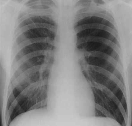
If someone experiences blunt or sharp trauma to their chest region, it’s important to consider the possibility of a traumatic pneumothorax, which is a type of lung injury. Doctors typically use patient history, a physical examination, and a chest X-ray to make a diagnosis. But, sometimes these methods can miss smaller pneumothoraces – a term for collapsed lungs. When that happens, a CT scan can help with diagnosis.
If a patient suddenly experiences sharp chest pain and difficulty breathing, it might be a spontaneous pneumothorax. This kind of lung collapse happens without any obvious trauma and should always be considered as a potential cause.
Usually, an upright chest X-ray can help doctors make a diagnosis, except in the case of a tension pneumothorax, which is typically diagnosed through clinical examination.
In many situations, doctors use point-of-care ultrasound to evaluate patients with a potential pneumothorax. Actually, an ultrasound can often spot pneumothoraces quicker than a standard chest X-ray, with the added benefit of not exposing the patient to any radiation.
Pneumothoraces can be small or large. A small pneumothorax shows a visible gap of less than 2 cm between the lung’s edge and the chest wall. A large pneumothorax has a gap of more than 2 cm. But be aware that a chest X-ray might make a pneumothorax look smaller than it actually is.
Treatment Options for Acute Pneumothorax Evaluation and Treatment
Treatment for pneumothorax, or lung collapse, depends on the cause, patient symptoms, and risk factors. The main goals of treating pneumothorax include removing the trapped air, reducing air leakage, healing the lung tissue, helping the lung to re-expand, and preventing any future occurrences.
People who have a pneumothorax but no symptoms may not need any immediate treatment, unless there’s a high risk of it happening again. However, this decision is usually taken outside of the emergency room, with a lung specialist (pulmonologist) deciding on the best course of action.
If someone has symptoms, but their vital signs – such as heart rate and blood pressure – are stable, they might need a needle or a small chest tube to remove the trapped air. Studies show that, particularly with a first-time pneumothorax, using a needle is as safe and effective as using a tube. These patients usually need to stay in the hospital to receive high flow oxygen and regular chest X-rays.
The majority of people with a pneumothorax caused by an injury (traumatic pneumothorax), whether their vital signs are stable or not, need a larger or smaller chest tube. A small tube is usually sufficient, unless the pneumothorax is very large, in which case a larger tube may be needed. If there’s also blood in the chest cavity (hemothorax), a large chest tube is definitely necessary.
Medication is mostly used for managing pain, either from the pneumothorax itself or from the procedures used to treat it. Pain relief may be achieved through local anesthetics applied around the site of the chest tube, intravenous and oral pain medication, or a combination of these. Chest tube insertion usually requires stronger pain relief medication, such as intravenous opiates or procedural sedation analgesia. Some experts also recommend regional anesthesia, such as intercostal nerve blocks. Preventive antibiotics may be used during chest tube insertion to avoid infection and further complications, like emphysema.
In cases of frequent pneumothoraces, a procedure called chemical pleurodesis, which involves sticking the lung to the chest wall with the use of talc, may be a treatment option.

lung.
What else can Acute Pneumothorax Evaluation and Treatment be?
When diagnosing a spontaneous pneumothorax (a collapsed lung that wasn’t caused by injury), doctors have to rule out other conditions that can cause similar symptoms. These include:
- Pneumonia (an infection of the lungs)
- An acute asthma attack
- Bronchitis (inflammation of the airways)
- A pulmonary embolism (a blood clot in the lungs)
- An aortic dissection (a tear in the heart’s main artery)
- Costochondritis (inflammation of the rib cage cartilage)
- Anxiety or panic attacks
- Injuries to the diaphragm (the muscle that helps you breathe)
- GERD (stomach acid that comes up into the esophagus)
- Mallory-Weiss Syndrome (a tear in the esophagus)
- Boerhaave’s syndrome (a rare condition where the esophagus ruptures)
- Mediastinitis (inflammation of the area between the lungs)
- Myocarditis (inflammation of the heart muscle)
- Pericarditis (inflammation of the sac around the heart)
- Pleurodynia (chest pain caused by a viral infection)
- Tuberculosis (a bacterial infection that mainly affects the lungs)
- Pulmonary empyema (pus in the space between the lung and chest wall)
- Lung abscess (a pocket of pus in the lungs)
In cases where a pneumothorax is caused by trauma, doctors must also consider the possibility of a tension pneumothorax (a life-threatening condition where air is trapped in the chest) or a hemothorax (bleeding in the chest). They also need to look for other injuries in the chest or abdomen, and a complete evaluation is necessary.
What to expect with Acute Pneumothorax Evaluation and Treatment
The likelihood of experiencing spontaneous pneumothorax (a condition where air gets trapped between the lung and the chest wall) again after the first episode is quite high – between 20 to 60% within the three years following the initial occurrence.
Possible Complications When Diagnosed with Acute Pneumothorax Evaluation and Treatment
Misdiagnosis is a common problem when dealing with pneumothorax, a condition involving air in the space surrounding the lungs. This can arise due to several reasons, including a partial or incomplete patient history, a not deeply investigated physical exam, not suspecting the condition, failing to get a chest x-ray, or missing the signs of pneumothorax when analyzing a chest X-ray. When pneumothorax is not correctly identified, it often results in the absence of needed treatment. In some instances, misdiagnosis can result in severe health complications such as:
- Mutation to tension pneumothorax, a severe form of the condition
- Hypoxemic Respiratory Failure, where there’s not enough oxygen in the blood
- Shock, a critical condition that can be life-threatening
- Respiratory arrest, which is when regular breathing pauses or completely stops
- Cardiac arrest, when the heart abruptly stops beating
- Empyema, a condition where pus accumulates in the space between the lungs and the inner surface of the chest wall
Additional complications may arise from the treatments for pneumothorax, which include needle decompression or thoracostomy. These complications include failure of the lung to re-expand, lung injury, infection at the procedure site, pleural space infection, blood vessel laceration, persistent air leak, damage to nerve and blood vessels bundle between the ribs, and possible irregular heartbeats caused by the chest tube. Further complications like air from the pneumothorax reaching the mediastinum, the cavity between the lungs, can also occur. This can be detected on a chest X-ray, seen as an abnormal lucency around the heart and could be associated with auscultation of abnormal heart sounds named Hamman’s crunch that is best heard when the patient is lying on their left side.


