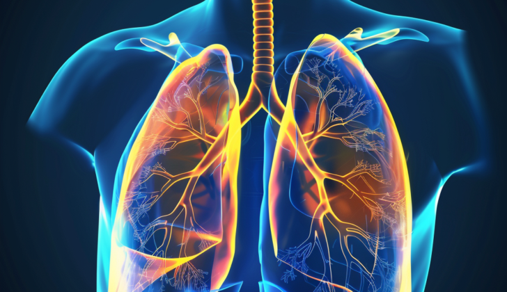
What is Pulmonary Interstitial Emphysema?
Pulmonary interstitial emphysema (PIE) is a rare lung condition that is most commonly found in newborns, but can also occur in adults. This condition happens when there is too much air pressure in the small air sacs of the lungs, which disturbs the surrounding lung tissue. This damage can lead to the formation of linear and cystic spaces in the lung, and air leakage. This escaped air gathers outside the normal airways and within the interstitium or bronchovascular complexes, affecting the lungs’ structure.
Premature babies suffering from PIE may experience a condition called respiratory distress syndrome. This condition is a serious concern as it can make it difficult for the baby to breathe efficiently and exchange gases. If not addressed, it can harm the lungs, lead to prolonged low oxygen levels, respiratory acidosis (an increase in acids in the body due to poor lung function), and pulmonary hypoperfusion (reduced blood flow to the lungs).
PIE is identified based on imaging tests and tissue examination. Treatments like surfactant administration (a substance that helps keep the air sacs in the lungs open) and high-frequency breathing support have helped reduce the occurrence of PIE in premature infants. The latest treatment for infants suffering from respiratory distress syndrome includes preventative treatment with synthetic surfactant and continuous positive airway pressure, with or without the use of a ventilator.
What Causes Pulmonary Interstitial Emphysema?
Possible causes of certain conditions in newborn babies include:
* Struggling to breathe normally or experiencing stress while breathing
* Being born too early or preterm
* Breathing in their first bowel movement (meconium)
* Use of intense or high-pressure artificial methods to aid in breathing
* Specific types of lung infection (like pneumonia or sepsis), the inhalation of fluid found in the womb, or infections in the placenta
* Incorrect placement of an artificial airway used for breathing support
* Exposure to a drug called Magnesium Sulfate before birth
In adults, similar conditions can be caused by:
* Asthma
* Smoking
* Physical injury to the lungs caused by rapid or excessive changes in air pressure
Risk Factors and Frequency for Pulmonary Interstitial Emphysema
PIE, or Pulmonary Interstitial Emphysema, often affects premature infants within the first weeks after birth. Babies who develop PIE within their first two days generally have a serious outlook. It’s common among infants with low birth weight, those born prematurely, and those affected by complications like lack of oxygen at birth and infections shortly after birth. Historically, it doesn’t seem to favor either gender. In fact, up to 24% of extremely premature infants, who were on breathing machines and received treatment to help their lungs develop, experienced PIE during their time in the hospital.
The number of cases differs depending on the group that’s being studied. For instance, one research found that 3% of newborns with breathing problems, who were born prematurely and treated with a lung developing drug early, developed PIE. Unfortunately, this number rose to 8% when the drug was administered late and soared to 25% in babies who weren’t treated with this drug.
An interesting study compared giving the lung developing drug as a preventative measure versus treating prematurely with it. The results showed more instances of PIE in premature babies and those who received the drug later in their treatment. To give more details, about 15% of the babies born 25 to 29 weeks early, who were treated with this drug right at birth had PIE, in comparison to 26% in the group of babies who didn’t receive the treatment.
Most cases of PIE occurred in premature babies that weighed less than 1000 grams at birth. The occurrences varied with the babies’ weights too: 42% for infants weighing 500 to 799 g, 29% for infants weighing 800 to 899 g, and 20% for those in the 900 to 999 g bracket.
Signs and Symptoms of Pulmonary Interstitial Emphysema
PIE, or pulmonary interstitial emphysema, is a condition that needs careful consideration and usually gets diagnosed with medical imaging and tissue examination called histopathology. There are some risk factors and signs that can suggest this condition. Common risk factors include premature birth and low weight at birth. People with PIE might show symptoms like needing more oxygen and having carbon dioxide build up quickly in their bodies.
It can be hard to diagnose PIE with just a physical examination because there are no distinct signs. But if PIE has progressed, there could be signs of air leaks which doctors will look out for. Symptoms of these air leaks can include things like faint breath sounds, a crackling sound when the affected area is touched, or a chest that looks bigger than normal.
- Common risk factors: premature birth, low birth weight
- Potential symptoms: need for more oxygen, rapid increase in carbon dioxide retention
- Signs of progressed PIE: air leaks causing faint breath sounds, crackling sound on the affected side, over-inflation of the chest
Testing for Pulmonary Interstitial Emphysema
PIE is a condition that can be partially diagnosed through medical imaging. When this is done, images of the lung tissue (also known as the parenchyma) typically appear filled with spherical cystic, linear, and oval, air-filled areas (lucencies). In the early stages, these lucencies appear as long, thin, linear structures but over time, they slowly progress to more round, cystic shapes. These linear lucencies can be about 3 – 8 mm long and less than 2 mm wide, while the cystic lucencies can measure from 1 – 4 mm in diameter.
During inhalation, the lungs might appear larger in size on imaging. However, in premature lungs, the tissue is less pliable which can result in the lungs appearing overly expanded on radiographs.
There might also be signs of air leaks visible on the imaging. These happen when the spaces around the lung tissue (interstitium) become filled with large volumes of air. This can cause disruptions in the gas exchange between the blood vessels and air sacs because the distance between them increases. Sometimes, this can lead to a condition called pneumothorax which occurs when air accumulates in the space that surrounds the lungs. This can compress the heart and disrupt blood flow to it. Other signs that might appear on imaging related to this condition include linear gas collections around the edge of the lung tissue. This usually happens when there is an increased need for respiratory support and bigger lung volumes, these can be informative signs of PIE.
Sometimes, early development of a condition called ‘bronchopulmonary dysplasia’ is seen along with partial blockages of the small airways (bronchia). Even in cases where PIE is not visible on imaging, it’s possible for autopsy studies to reveal its presence in infants with this condition. There was a case study that showed a CT scan taken before an autopsy exhibited thickened pleural wall and narrowed airways. The imaging also showed that the lungs were excessively inflated (hyperinflated) and that there was localized emphysema (damage to the air sacs in the lungs).
PIE can sometimes be detected using a conventional chest X-ray taken from the front while the patient lays flat on their back, but it often requires multiple scans over a period of time to track the progression of the disease. Identifying the difference between over-inflated small airways (lucent bronchioles) and PIE can sometimes be challenging. However, it is important to note that over-distended airways are typically round and uniform, unlike the irregularities seen with PIE.
In some cases, PIE can be misinterpreted as other lung conditions such as pulmonary edema or aspiration syndrome, especially if the normally aerated lung tissue appears to be surrounded by fluid leakage. If an infant is using a mechanical ventilator, the airways and air sacs (alveoli) might seem similarly enlarged as with PIE. If a chest x-ray doesn’t conclusively differentiate between these conditions, the next step would likely be the use of a CT scan for further evaluation.
Treatment Options for Pulmonary Interstitial Emphysema
Babies with PIE, or lung disease, need to be treated in a neonatal ICU. This illness can become critical and lead to complications like pneumothorax (collapsed lung) or pneumopericardium (air trapped around the heart), requiring the baby to be put on a ventilator. Other procedures may be necessary, too, like thoracentesis – removing fluid from around the lungs. The best way to manage PIE varies. A lot of babies with PIE are premature.
There are some tactics that could lessen the risk of PIE in premature babies. Using a treatment called surfactants in preemies less than 30 to 32 weeks old could help prevent PIE from developing by averting respiratory distress syndrome. It’s also beneficial to avoid using mechanical ventilation on the baby if possible, as high oxygen pressure can harm their undeveloped lungs. Instead, a technique called CPAP (continuous positive airway pressure) should be tried first, but it still carries a risk for causing PIE. If ventilation must be used, certain settings should be aimed for. The goal should be to have a short inhalation time, long exhalation time, and less pressure when adjusting a setting called PEEP. This allows for a proper emptying of the airway after exhaling. Monitoring is also necessary – checking vitals, the baby’s oxygen, blood gases, and nutrition is crucial.
There are ways to try and avoid PIE. Premature babies at risk of respiratory failure can be given a preventative dose of surfactant, which can decrease their risk. The occurrence of PIE lowers in premature babies if the surfactant is given early on with brief ventilation.
In the case of babies with PIE only on one side, a method called lateral decubitus positioning can be used. This approach positions the baby on their side and could resolve one-sided PIE in 2 to 6 days. It has a low failure rate and PIE rarely comes back. This positioning also helps with PIE on both sides if one lung is significantly more affected than the other. The healthier lung will get more oxygen, reducing the need for ventilation.
Surfactant is used in premature babies to prevent respiratory distress syndrome. It is administered into the throat and works by stopping the air sacs in the lungs from collapsing. There are natural surfactants, which come from animals and are similar to human surfactants, and synthetic ones. Some advantages of synthetic surfactants are that they are therapeutic and sometimes more effective than natural surfactants.
High-frequency positive pressure ventilation can prevent air leaks and PIE, but the evidence is limited. For babies in severe distress, selective intubation of the clear bronchus, or breathing tubes, can be effective. Some settings are better for treating PIE in babies, such as high-frequency oscillatory ventilation, which involves the rapid oscillation of the airway pressure. However, these settings need to be monitored as they can cause the trapping of gas and airway collapse.
If medical treatments don’t work and PIE doesn’t resolve on its own, a lobectomy, or surgery to remove part of the lung, may be needed. While this is a last resort, it is an option for infants who have serious lung disease.
Other less common treatment methods include artificial pneumothorax (using air or gas to make the lung collapse), chest physiotherapy with oxygen therapy, steroid treatment, ECMO (extracorporeal membrane oxygenation, or lung bypass), and nitric oxide treatment. Each of these is rarely used.
What else can Pulmonary Interstitial Emphysema be?
When a doctor is trying to diagnose PIE, they are relying on images and tissue samples. There are several conditions that might look like PIE at first, but can usually be distinguished using a type of x-ray known as a CT scan. These conditions include:
- Pulmonary edema (excess fluid in the lungs)
- Pulmonary embolism (a blood clot in the lungs)
- Bronchogenic cysts (cysts that develop in the airways)
- Congenital lobar emphysema (a lung abnormality present at birth)
- Air bronchograms in respiratory distress syndrome (abnormal air patterns in a lung disease often seen in premature infants)
- Aspiration pneumonia (a lung infection caused by inhaling material into the lungs)
- Diaphragmatic hernias (an opening in the diaphragm, the muscle that helps us breathe)
- Congenital cystic adenomatoid malformation (a rare lung condition present at birth)
The doctor must carefully consider and rule out these conditions in order to correctly diagnose PIE.
What to expect with Pulmonary Interstitial Emphysema
The diagnosis of PIE (Pulmonary Interstitial Emphysema) usually points to a serious health situation. Research has indicated high death rates of 53% to 67%. The outlook is particularly severe for babies born with low weights of less than 1600 grams and those with serious respiratory distress syndrome, with a noted death rate of 80%.
PIE can cause air leaks in the chest, such as pneumothorax and mediastinal emphysema, which further increase the risk of death. If PIE develops early, it’s associated with a higher risk of death due to serious underlying lung disease.
Proper management can help resolve PIE over several weeks. However, this may result in a longer dependence on a mechanical ventilator, leading to complications like bronchopulmonary dysplasia or chronic lobar emphysema. Occasionally, these conditions might necessitate surgical lung removal.
One study found that 54% of babies who survived PIE developed chronic lung emphysema, and half of these babies needed surgical lung removal. Research also indicates that babies with PIE are likely to develop brain bleeding and are still at a high risk of death.
Possible Complications When Diagnosed with Pulmonary Interstitial Emphysema
PIE, also known as Pulmonary Interstitial Emphysema, is a serious disease. There are various complications that can arise from it, including:
- Respiratory insufficiency – difficulty in breathing
- Mediastinal emphysema – air trapped in the middle part of the chest
- Other types of air leaks, such as pneumomediastinum (air trapped around the lungs), pneumothorax (collapsed lung), pneumopericardium (air around the heart), pneumoperitoneum (air in the abdominal cavity), and subcutaneous emphysema (air under the skin)
- Intraventricular hemorrhage – bleeding into the brain’s ventricular system
- Massive air embolism – air bubbles in the bloodstream that can block it
- Chronic lung disease of prematurity
- Periventricular leukomalacia – damage to the brain’s white matter near the ventricles
- Death
Recovery from Pulmonary Interstitial Emphysema
It’s essential to keep a special watch on premature babies after their treatment. Some of the complications to look out for include damage to the brain’s white matter (periventricular leukomalacia), bleeding in the brain (intraventricular hemorrhage), and delays in their development. These infants could also develop long-term lung conditions, requiring extended pulmonary care.
At present, medical research is undecided about whether treatment with drugs that open up the airways (bronchodilators) can help premature infants who have developed long-term lung disease.
Preventing Pulmonary Interstitial Emphysema
It’s crucial to talk about ways to prevent potential complications during pregnancy, especially for babies who are at a high risk for PIE. Here are some key points to remember:
Firstly, avoid smoking. Secondly, recreational drug use, such as cocaine and marijuana, must be avoided. Alcohol use should also be completely avoided. Lastly, it’s important to ensure proper prenatal care throughout the pregnancy.


