What is Foot Drop?
Foot drop is a condition where a person is unable to lift the front part of their foot due to weak muscles responsible for this action. This can lead to an abnormal, pain-avoiding way of walking that could potentially result in trips and falls.
The reasons behind foot drop can vary widely. These can range from problems with muscles, nerves, the spine, or autoimmune and musculoskeletal disorders. Depending on the specific cause, the strategies used to treat foot drop will differ.
Before a treatment can be determined, it’s necessary to understand what’s causing the issue. Here we will discuss the causes, symptoms, diagnosis, and treatment strategies for foot drop.
The nerves and spinal features involved in foot drop include:
Lumbar Nerve Roots: These are nerve roots that come out from the area where two backbone bones (vertebrae) touch each other. The L5 nerve root, important in foot drop, comes from between the L5 and S1 vertebrae.
Lumbar Plexus: This is a network of nerves (from L1 to L4 vertebrae) in the lumbar region. Several nerves important for muscle functioning emerge from this network.
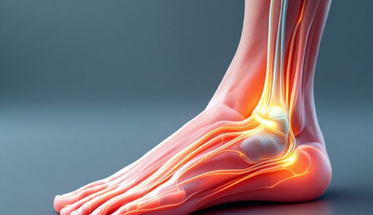
Sciatic Nerve: The sciatic nerve is the largest nerve arising from nerves L4 to S4. It controls muscles in the back of the thigh and keeps traveling into the back of the leg into a space called the popliteal fossa, where it splits into two: the tibial and common fibular nerves. The tibial nerve controls muscles that help to bend the foot and toes downwards.
Common Fibular Nerve: This is a branch of the sciatic nerve that moves towards the side of the calf muscle and reaches the fibular bone. This nerve is prone to compression injuries. It further divides into deep and superficial nerves. The deep fibular nerve controls foot and toe upwards movement and provides sensation to the area between the first two toes. The superficial nerve controls outward twisting of the foot and provides sensation to the top of the foot and side of the calf.
These nerves and muscles play an important role in managing foot drop, and understanding their function can help to navigate the diagnosis and treatment of this condition.
What Causes Foot Drop?
The fibular nerve, which is located in your leg, can sometimes get trapped or squeezed along its pathway, causing a condition known as compressive neuropathy. One common type of this condition occurs when the fibular nerve is trapped at the fibular head, an area towards the top of your lower leg bone (the fibula). This is especially likely when a muscle in the same area, the biceps femoris, grows in a way that forms a tunnel structure and traps the nerve. Other things that can contribute to this include weight loss, spending a long time in bed, wearing tight casts, and certain illnesses that cause growths or affect your bones.
Something similar can happen with the sciatic nerve, located in the back of your lower body. For example, this nerve can be squeezed between two parts of a muscle in your buttocks, leading to a condition known as foot drop, where you have issues lifting the front part of your foot.
If you spend a long time in an Intensive Care Unit (ICU), this can lead to other types of nerve problems. Almost 1 in 10 patients spending more than 4 weeks in an ICU develop weakness, or paresis, of the fibular nerve. People with diabetes are especially vulnerable to these nerve issues.
Another cause of foot drop can be a condition called lumbar radiculopathy, which happens when nerves in the lower back are squeezed or irritated. Typically, this can happen when the discs in the spine slip out of place or become swollen.
Orthopedic injuries, like bone dislocations, fractures or blunt trauma can also cause nerve problems that result in foot drop. For example, injury to the hip or complications from surgery can damage the sciatic nerve, leading to issues like foot drop.
A number of neurological conditions can also result in foot drop. These include Amyotrophic Lateral Sclerosis (ALS) also known as Lou Gehrig’s disease, which causes motor neuron cells in your body to die, leading to muscle weakness. Cerebrovascular diseases (illnesses related to blood circulation in the brain), can cause one side of the body to become paralyzed (hemiplegia), and foot drop is a possible part of this.
Conditions such as AIDS, leprosy, hepatitis, among others can affect individual sensory and motor nerves, disrupting nerve function and eventually leading to muscle weakness which can include foot drop. Guillain-Barré syndrome is an autoimmune disease that results in progressive motor weakness and sensory loss; foot drop can be a symptom here as well. Charcot-Marie Tooth (CMT) disease is a common inherited nerve disease where one of the main symptoms is foot drop. Last but not the least, disturbances of the mind (like somatization disorder and conversion reaction) can also produce symptoms that resemble foot drop.
Additionally trauma can lead to build up of pressure in a specific compartments of the leg, that can restrict blood supply to the peroneal nerve leading to foot drop. There are also numerous medical procedures or factors related to prolonged bed rest that can accidentally lead to foot drop.
Risk Factors and Frequency for Foot Drop
Fibular neuropathy affects about 19 in every 100,000 people and is more common in men than women. After a total knee replacement, the rate is reported to be 0.79, with slightly more men affected than women at a ratio of 2.8 to 1. Most of the time, it only affects one leg, with the right and left sides being equally likely to have the condition.
The condition known as ALS affects around 1.54 in every 100,000 people each year globally. It can occur at any age, but is common between 50 and 75.
Unfortunately, the frequency of mononeuritis multiplex in both the United States and the rest of the world isn’t known.
- Fibular neuropathy incidence is about 19 per 100,000, with males more affected than females.
- The ratio of males to females impacted by fibular neuropathy after a total knee replacement is 2.8 to 1.
- The condition usually affects only one leg, with no preference for the right or left side.
- ALS is estimated to affect 1.54 per 100,000 people globally each year and usually occurs between ages 50 and 75.
- The frequency rate for mononeuritis multiplex is unknown.
- The annual occurrence for AIDP ranges between 1.0 to 1.2 per 100,000 people and men are about 1.5 times more likely to be affected than women.
Signs and Symptoms of Foot Drop
Investigating foot drop involves both a careful review of the patient’s medical history and a physical examination. The exam is geared towards identifying the particular nature of the problem, with some specific tests being applied.
Musculoskeletal testing usually takes the form of having the patient perform certain movements, such as standing on their toes, standing on their heels, and bending their knees deeply. A health professional will also rate the power of major muscle groups in the lower body on a scale of 0 to 5. These muscles include those responsible for ankle movements and bending and straightening of the knee and hip. A sensory examination for pinpricks is also carried out in the areas of skin supplied by specific peripheral nerves and parts of the skin supplied by nerves from the lower back.
A comparison of muscle mass between the two sides can also provide useful information. This can involve making measurements around various parts of the legs and noting any differences. Using these measurements, any changes in muscle mass over time can be tracked. However, this doesn’t typically include a detailed assessment of specific sensory nerves.
Further testing, such as an electrodiagnostic consultation, could be necessary. This involves tests of muscle activity and nerve function, and it often requires input from a specialist. This specialist often isn’t the first healthcare professional involved in the patient’s care.
Foot drop can be caused by a variety of issues. These could range from damage to the spinal roots that are linked to the nerve responsible for foot movement, to damage to the nerve itself.
Damage to areas such as the L5 root, lumbar plexus, sciatic nerve, and deeper nerves can lead to weakness in the muscles at the front of the lower leg. This is due to these muscles’ role in lifting the foot, and this weakness can result in foot drop. The presenting symptom is a decrease in the ability to walk compared to before. Other signs may include inability to raise the foot during the initial contact with the ground, foot dragging, or tripping over the foot.
Foot drop can also be caused by issues such as issues with the fifth lumbar nerve root, problems with the lumbosacral plexus, or neuropathy of the sciatic nerve, common fibular nerve, and other surrounding nerves. These present with pain or numbness in specific areas of the leg and foot and can involve weakness of specific muscles.
Foot drop can significantly affect the way a person walks. Normally, walking involves alternating between having one foot on the ground while the other is in the air. The cycle begins with the heel of one foot contacting the ground and ends when the same heel contacts the ground again. The foot is usually flat on the ground while the body’s weight is on it and is then raised off the ground in anticipation of the next step. In foot drop, the foot often remains pointing down while on the ground, making it harder to clear the foot from the ground for the next step. This can result in tripping or the need to lift the foot higher than usual.
Specific muscles should be evaluated for any weakness that could result in foot drop. These include the hamstring muscles, muscles moving the toes and ankle, muscles at the back of the lower leg, hip adductor muscles, among others. Assessing the strength of these muscles can be crucial in diagnosing and treating foot drop.
Testing for Foot Drop
After a detailed physical check-up, your doctor will likely suggest X-rays of your pelvis, tibia (shin bone), and fibula (calf bone) to make sure there are no fractures or dislocations. If they suspect nerve injuries due to lumps or tumors, they might suggest a magnetic resonance imaging (MRI) test. An MRI can provide detailed pictures of the lumbar spine (lower back), knee, and ankle, and can help identify any potential soft tissue lumps that might be compressing your nerves. Modern, advanced MRI techniques can also provide information about the nerve itself, such as any changes in shape, size, or overall health.
Apart from these images, ultrasound (a technology that uses sound waves to create images of the inside of your body) can also show swelling around or near the compressed nerve.
Concerning blood tests, the doctor might order tests to look for rheumatoid factors and ANA (antinuclear antibodies), which could indicate autoimmune conditions. A complete blood count (CBC) test and a chemistry panel, which are comprehensive blood tests, might also be carried out to rule out other causes of your symptoms.
Electrodiagnostic studies can confirm the diagnosis and provide information about the severity of your condition, as well as give some insights into the expected recovery time. This type of investigation is composed of two parts:
1. Nerve conduction studies: These tests provide information about the health of your nerves’ protective layer, the myelin. If a nerve is compressed, this test will show a delay in the nerve’s signal transmission.
2. Needle electromyography (EMG): This test involves inserting tiny needles into muscles that are connected to the nerve being studied. The doctor looks at the nerve signals in these muscles while you contract them. The presence of certain electrical signals called positive sharp waves and fibrillation potentials indicates nerve damage.
In an EMG, at least one muscle connected to the superficial peroneal nerve (a nerve in your leg) and two muscles linked to the deep peroneal nerve are usually examined. The test can also be performed on muscle connected to the tibial nerve (a nerve running down the inner part of your leg).
These tests help identify the location and severity of the lesion (injury or damage) and estimate chances of recovery. If the tests find no signs of nerve damage and the nerves in the muscles are working well, the chances of a good recovery are high. On the other hand, if there are signs of nerve damage and the nerves in the muscles are not working well, the chances of a full recovery are less likely.
Sometimes health conditions related to the immune system and severe illnesses can cause nerve or muscle problems. Often, after a thorough check-up, the doctor might diagnose a “clinical syndrome” which describes a group of symptoms rather than a single clear-cut condition.
Treatment Options for Foot Drop
The approach to the patient’s care largely depends on what is causing the foot drop and the nature of any damage or compression. After evaluating and diagnosing the patient, many treatment options may be considered.
Surgical Options:
For patients who have experienced some form of trauma that has led to nerve damage, nerve reconstruction surgery is usually performed. This surgery should ideally take place within 72 hours of the injury.
If the nerve is being compressed, a surgical procedure to remove the decompressed tissue and free the nerve may be needed. This procedure has shown a high success rate, with about 97% of patients regaining function. However, depending on the extent of the nerve injury, additional procedures like nerve reconstruction using a graft, or nerve conduits might be necessary. Patients may also need a surgical release procedure if they have developed an equinus deformity, a condition affects the angle of the ankle.
In cases where the nerve dysfunction is quite severe, nerve or tendon transfers may be required.
However, for other causes of foot drop, treatment initially tends to be conservative, because there is a possibility that the symptoms may improve spontaneously over time.
Conservative Management:
Conservative management includes physical therapy, splinting, and medication for pain relief. The primary goal of this approach is to stabilize the patient’s gait, and prevent accidents like falls and muscle contractures. Physical therapy mainly aims to stretch and strengthen the muscles, and electrical stimulation techniques also show promise in treating weakened muscles. The patient is encouraged to perform home exercises to maintain muscle strength and flexibility, and prevent muscle contractures.
Splints like an ankle-foot orthosis (AFO) are used to help prevent further complications, especially in more serious nerve palsies that aren’t showing signs of recovery. Patients receive education on how to use and care for these devices.
Special instructions for skin care are vital for patients with numbness to avoid skin abrasions and ulcers. This care is usually coordinated with the professional creating the AFO.
Medications used for pain relief include topical analgesics, serotonin reuptake inhibitors, membrane stabilizers, and opioids. It’s worth noting that while the medication may relieve pain, it won’t necessarily result in a recovery of nerve function.
Follow-up checks, which may include a reassessment of the condition and testing for nerve regeneration, are an integral part of the treatment plan.
Surgical Management:
Depending on the specifics of the patient’s condition, several different types of surgeries may be considered:
– Nerve repair: This may involve direct primary repair if the nerve damage is minor; a nerve conduit if the gap in the nerve is less than 3 cm; or an autologous nerve graft if the gap is significant.
– Neurolysis and nerve decompression: These procedures are done to relieve pressure at certain areas causing compression, such as the fibular tunnel or fibular head.
– Nerve or tendon transfer: This can be done to restore the ability to flex the ankle with minimal side effects.
If the nerve transfer isn’t effective, the patient may undergo tendon transfer surgery, with the posterior tibial tendon being the most common choice.
In difficult cases that don’t respond to other treatments, the patient may undergo ankle fusion surgery. The ankle is fixed in a 90° position relative to the tibia through either a pantalar (involving the talocalcaneal, talonavicular, and tibiotalar joints) or a tibiotalar fusion.
What else can Foot Drop be?
When someone has a stroke, it may lead to weakness in the entire body part, often making the limb seem artificially long. The patient may then walk by making a circular motion with their leg, a movement called circumduction. They may also find it hard to swallow, speak or move their arm.
Impairments in the cerebellum, a part of the brain that controls smoothness and balance of movements, could cause imbalance while walking. This is known as a “cerebellar gait”, where the person finds it difficult to walk straight.
Another type of difficulty with walking is called “ataxic gait”, where both sides are affected. This type of walking issue arises when long tracks of the spinal cord are affected, causing loss of position and vibration sense. It often results in high steps and a side-to-side sway, like in individuals suffering from alcohol use disorder.
Severe radiculopathy (nerve root inflammation) in the lower back (L5 level) can also cause gait disturbances.
In Parkinson’s disease, a part of the brain called the substantia nigra is affected. This leads to issues with walking, with problems starting to walk and taking quick, short steps, a condition called “festinate gait”.
Issues with the network of nerves in the lower back (lumbar plexus) like a compressive tumor can also affect the way a person walks. Diabetic amyotrophy, a nerve disorder associated with diabetes, can also cause similar problems.
Finding no physical cause for walking problems may suggest psychological factors. Some psychological disorders like conversion reaction, somatization disorder, and malingering could cause gait disturbances. Other mental health problems like depression and anxiety should also be considered when all physical evaluations are normal.
Surgical Treatment of Foot Drop
When there’s a growth or a mass in the body that takes up space (known as a space-occupying lesion), a person may experience weakness in their leg. This happens when the growth impacts the network of nerves in your lower back area, known as the lumbosacral plexus. So, if someone is encountering leg weakness without a clear reason, along with other general signs of health problems, it’s important to consider if a cancerous growth could be the root cause.
Nonetheless, it’s more commonly seen that this type of growth affects specific nerves in the leg resulting in a condition known as genital femoral neuropathy. This can lead to weakness in the ability to straighten the knee. So, if you’re facing unexplained weakness, especially when trying to extend your knee, it’s important to consult your doctor to explore the possibility of an underlying health issue.
What to expect with Foot Drop
The outcome of an injury can depend on the severity of the damage and what caused it in the first place.
If you’ve experienced Neurapraxia, a kind of temporary damage to your nerves, it’s usually the case that you get back to normal in about 3 months. This is because your nervous system is able to recover relatively quickly.
However, if you’ve had axonal loss, this means some nerve fibers have been damaged. In this case, recovery might take longer, somewhere between 6 to 12 months.
Then there’s Neurotmesis, which is the most severe type of nerve damage. The prognosis here is quite uncertain and often you might require surgical treatment, like getting a nerve or tendon transferred. The best time for a nerve surgeon to take a look at your injury and possibly perform a procedure would be around 6 to 9 months after the injury has occurred.
Something to keep in mind is that if a muscle has been without nerve stimulation for over 18 months, it might not regain much functionality, even after treatments.
If the injury was caused by differing factors like ALS (a nerve disease) or space-occupying lesions (lumps or growths), the outcome will depend more on tackling these specific conditions rather than just focusing on the nerve damage itself.
Possible Complications When Diagnosed with Foot Drop
Some complications may come directly from nerve damage, while others may stem from the use of walking aids and braces.
One problem that could occur from nerve damage is “foot drop,” which hinders a person’s ability to lift the foot off the ground and may cause stumbling or falling. The use of walking aids like walkers and canes can also lead to issues, especially when moving on uneven grounds. Other complications involve the skin: when it is numbed, it can lead to ulcers, and ill-fitting braces might cause physical wear and abrasions. Depending on the changes in a person’s body size, adjusting the brace might be necessary. In certain conditions such as kidney failure and congestive heart failure, changes in the body’s fluid levels can cause swelling; different braces may be needed to accommodate these changes.
Complication Summary:
- Nerve damage leading to foot drop, causing falls
- Difficulty using walking aids such as canes and walkers on uneven surfaces
- Ulcers from numbed skin
- Abrasions from poor-fitting braces
- Braces may need to be adjusted based on body size changes
- Swelling conditions like kidney and congestive heart failure may require different braces
Recovery from Foot Drop
As mentioned earlier, physical and occupational therapy can make a big difference in a patient’s treatment. These therapies can influence whether a patient gets treated through non-invasive methods or needs to undergo surgery.
Preventing Foot Drop
For successful treatment, it’s crucial that the patient is involved in each step of the decision-making process. They, along with their family members, should receive education about their illness. This includes understanding the cause of the disease, how it’s diagnosed, its severity, and how it affects the body. Helpful handouts and specific guidelines may discuss at-home exercises, skincare routines, and how to care for any braces they may need.
The healthcare provider must have the ability to communicate with the brace expert even between medical visits. This is important because all braces wear out eventually and need to be replaced or repaired. The patient should also receive clear instructions for any future check-ups. This ensures that any changes in treatment, brace wear, or need for more diagnostic tests can be properly tracked and managed.


