What is Neurothekeoma?
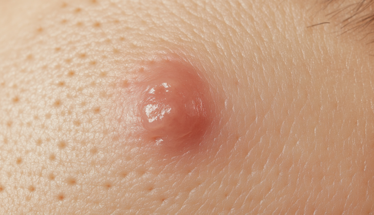
Neurothekeomas are uncommon, non-cancerous, surface-level lumps, most often found on the head and neck. This condition usually affects women more than men, primarily seen in their twenties and early thirties. Neurothekeomas usually appear as a skin-colored, pink, red, or brown lump that is clearly outlined. Most of the time, they don’t cause any symptoms but can sometimes be painful if pressed.
These lumps can look differently under a microscope depending on how much of a certain substance called the myxoid matrix is present. This has led to them being categorized into myxoid, cellular, or mixed types. In the past, due to similarities in how they look and where they occur, it was thought that neurothekeomas could be a type of nerve sheath myxoma, another kind of benign tumor.
However, research by Barnhill and Mihm in 1990, suggested that neurothekeomas are actually a different problem altogether. Instead of originating from the protective covering of nerves (nerve sheath), it’s thought that neurothekeomas arise from a type of cell known as a fibrohistiocyte.
While nerve sheath myxoma reacts with a certain protein called S100 when tested, neurothekeoma does not, regardless of whether it is a myxoid, mixed, or cellular type. This further differentiates neurothekeoma from nerve sheath myxoma.
What Causes Neurothekeoma?
There’s been a lot of confusion when it comes to understanding and defining neurothekeomas. In 1980, researchers Gallager and Helwig first classified neurothekeomas as skin tumors that originated from nerves. They said these tumors typically appeared as flesh-colored or slightly red nodules that were soft to touch. These nodules were found on the head, upper limbs, or body, and were more common in young women.
Under a microscope, these tumors showed thread-like cells arranged in groups with a jelly-like background containing skin collagen bundles. Since the tumor cells looked similar to Schwann cells (cells that form a protective covering of nerves) under an electron microscope, the researchers named them “neurothekeoma”, derived from the Greek word for sheath.
In 1986, another researcher, Rosati, reported cases of cellular neurothekeoma, believing them to be a sub-type of the previously identified neurothekeoma. In 1990, Barnhill and Mihm studied several cases of cellular neurothekeoma. Their findings suggested that cellular neurothekeoma was distinctly different from the original reported version of neurothekeoma. By 1991, they suggested that cellular neurothekeoma may be a completely separate type of tumor.
It is now accepted that some of the cases initially reported as neurothekeoma in 1980 and some subsequent cases were actually a different type of tumor called dermal nerve sheath myxoma. This type of tumor has similar characteristics to the neurothekeoma but originates from the peripheral nerve sheath (the outer covering of nerves). The confusion arose due to overlapping similarities in clinical appearance and features under the microscope.
In 2005, Fetsch and his team studied a series of nerve sheath myxomas, discovered their characteristic immune profile, and found that these tumors tend to recur more often than neurothekeomas. Further research was done in 2007 which presented a more detailed profile of neurothekeomas by defining them as distinctly different from nerve sheath myxoma. It’s been suggested that neurothekeoma may not originate from nerve coverings like initially thought, but may be of a fibrohistiocytic origin, meaning it might originate from cells that are associated with the formation of connective tissue and immunity.
Risk Factors and Frequency for Neurothekeoma
When looking at the numbers, women are twice as likely as men to get this particular type of tumor. It can occur at any age, but it is most commonly seen in people in their 20s and 30s.
Signs and Symptoms of Neurothekeoma
Neurothekeomas, or lesions, usually appear as a single, small lump on the skin. These bumps are less than 2 cm in size and might be skin-colored, pink to tan, or red to brown. These lumps on the skin generally don’t cause any symptoms, but can be painful to touch. They grow slowly and usually only affect the surface layers of skin. Rarely, they might affect deeper layers of skin or even the muscle underneath. Neurothekeomas are most commonly found on the head and neck but can also show up on the shoulder or upper arms. It is rare to find these lesions on the mucous membranes, which are the soft, moist areas inside your body, like inside your mouth or nose.
Testing for Neurothekeoma
For the proper diagnosis of a medical condition known as neurothekeoma, it is essential to pair the studies of disease (clinical) with the studies of disease in tissues (pathological). This disease often shows up as skin nodules that could potentially be deeper than just the skin’s surface. If the nodule is very shallow, a shave biopsy might be performed. This is a simple procedure where a sample of the nodule is shaved off for further examination.
However, given the possibility that these nodules might extend deeper into the skin, other types of skin samples may be needed at times. These could be a deep punch biopsy, which is a circular incision; or an excisional biopsy, where the whole nodule and some of the surrounding tissue is removed. The type of skin biopsy chosen depends on the size and location of the nodule.
If the nodule is small, easy to feel and is not located in an area that is either sensitive or visible, imaging tests like CT scans or MRIs are generally not necessary. However, if effort is required for surgical planning or further understanding, imaging techniques like Computed Tomography (CT), Magnetic Resonance (MR), or a kind of imaging called FDG PET (which uses a radio-active drug to create images of the body) might be helpful.
Despite these technological aids, the ultimate method for diagnosing neurothekeoma continues to be histology, an examination of the tissues under a microscope.
Treatment Options for Neurothekeoma
Surgery is usually the most effective treatment for removing neurothekeomas, which are usually small, benign (non-cancerous) tumors. Doctors strive to remove the tumor fully, aiming for what’s called “clear margins”. This means they try to take out a little bit of normal tissue around the tumor to make sure no tumor cells are left behind. The aim is to remove all of it without affecting nearby healthy cells.
If the tumor is in a visually prominent place, like the face, a specific type of surgery called Mohs micrographic surgery might be used. This approach is designed to achieve the same goal — complete tumor removal — but with a minimal impact on the surrounding normal tissue to preserve the patient’s appearance.
While most of these tumors are small (less than 1 cm in size) and are not cancerous, some cases have shown features that doctors might find concerning. These features include tumors larger than 1 cm, unusual cell shapes and sizes (called pleomorphism), increased cell division (mitotic activity), extension into the muscle or under-the-skin fat, and invading blood vessels.
But even with these unusual features, the chances of the tumor coming back after complete surgical removal are quite low. This means that once it’s removed, it usually does not return.
What else can Neurothekeoma be?
When doctors try to determine what is causing an abnormal mass in the head and neck region, they consider a number of possibilities. These can include:
- Granular cell tumor
- Cellular neurothekeoma
- Nerve sheath myxoma
- Neurofibroma
- Schwannoma
- Benign fibrous histiocytoma
- Melanocytic lesions, for example, Spitz nevus or melanoma
- Infection
If doctors don’t find any bacterial or fungal elements, this means that an infection is unlikely to be the cause. One way doctors can rule out a granular cell tumor is if a test for S100 protein, which this type of tumor typically has, is negative.
Other conditions on the list, like neurofibromas, schwannomas, and nerve sheath myxomas, fibrohistiocytic neoplasms like neurothekeoma and fibrous histiocytoma, or melanocytic lesions, can appear similarly to solitary tumors and may also imitate each other.
Identifying between neural and fibrohistiocytic masses and cancerous tumors like melanoma can be difficult due to the various appearances of epithelioid cells and atypical features. However, details of their configurations can help differentiate fibrohistiocytic masses from each other. Detecting at least some areas of nested growth, for instance, can help identify a cellular neurothekeoma, which would look differently than superficial angiomyxomas or benign fibrous histiocytomas.
Differentiating between cellular neurothekeoma and nerve sheath myxoma can be challenging due to their similar appearances. But tests known as immunohistochemistry can help distinguish these. For example, nerve sheath myxomas are consistently positive for S100 and negative for NKI-C3, while cellular neurothekeomas show a different pattern. The remaining neural tumors on the list can also be distinguished through their morphology and the results of similar tests.
What to expect with Neurothekeoma
Neurothekeoma is deemed to be a benign or non-dangerous condition. As a result, people with this condition have a great probability of recovery. It also tends to come back very rarely after the entire tumor has been surgically removed.
Possible Complications When Diagnosed with Neurothekeoma
Most of the time, complications after removal of neurothekeomas, a type of skin tumor, are minor and only affect the appearance of the surgical scar. However, if the tumor has penetrated beyond the layer of fat below the skin and into the muscles, the surgery can lead to localized weakness due to the amount of muscle that had to be removed.
Common complications:
- Surgical scar affecting appearance
- Local weakness in muscle if tumor penetrated deeper layers and muscle removal was necessary
Preventing Neurothekeoma
The cause of neurothekeoma, a rare type of skin tumor, is still not known. Therefore, at present, doctors are unable to provide advice on how to prevent these skin growths.


