What is Mandible Body Fracture?
Fractures of the jawbone make up about 25% of all facial bone breaks. Out of all the jawbone breaks, 11% to 36% occur in the main part of the jawbone. The main cause of these injuries is usually physical violence.
What Causes Mandible Body Fracture?
The reason someone might experience a body fracture often involves incidents like personal assault, car accidents, falls, or accidents during sports activities. The most frequent reason is car accidents, which make up 43% of cases. They’re followed by assaults, which account for 34% of fractures. Falls make up 7% of fracture cases, and sports accidents are the cause in 4% of instances.
Risk Factors and Frequency for Mandible Body Fracture
Research has shown that out of all fractures in the lower jaw, nearly 29% are fractures of the body of the jaw. These are most commonly observed, followed by fractures of the condyle and angle of the jaw. For children, fractures of the jaw’s condyle and body are the most common facial fractures. Additionally, fractures of the body of the jaw occur more often in males compared to females.
- Nearly 29% of all lower jaw fractures happen in the body of the jaw.
- The next most common areas for these fractures are the condyle and angle.
- In children, the most common facial fractures occur in the body and condyle of the jaw.
- Jaw body fractures are more common in males than in females.
Signs and Symptoms of Mandible Body Fracture
When evaluating someone who may have fractured their jawbone, several factors need to be considered. You need a thorough background information about the patient. This includes preexisting conditions such as bone diseases, cancer that affects bone, arthritis, collagen vascular disorders, and issues with the jaw joint. The nature of the injury, including the type and direction of the force that caused it, can help determine whether additional fractures may be present. For example, car accidents often involve a much larger force and can result in numerous, complex fractures. On the other hand, violence-related injuries might result in a simple, stable fracture.
In examining the patient, you may notice changes inside the mouth. A change in how the teeth fit together or sensations like numbness or tingling in the lower lip might become apparent. These symptoms usually occur when fractures are located further back along the jaw and are often not seen in stable jaw fractures.
Telltale signs of a fracture, in general, include a cut in the gum tissue, misaligned teeth, and/or bruises inside the mouth. A fracture’s movement can be checked by gently handling the jaw and applying pressure between the top and bottom teeth.
Changes can also be noticeable on the outside of the face. Facial contour might change due to a loss of jaw structure or skin abrasion. A fracture can result in a flattened appearance of the side of the face. When there’s a loss of the jaw structure, a serious fracture should be considered. The front of the face may be moved forward, leading to an elongated appearance. In these cases, the lower jaw may also be displaced downward. Other potential signs include cuts, swelling, and bruising.
Testing for Mandible Body Fracture
To evaluate fractures, doctors often use radiographs and CT scans. These can involve different views, like panoramic, lateral-oblique, posteroanterior, and more. Each offers a unique perspective for diagnosing different aspects. For instance, the lateral-oblique view is helpful for diagnosing fractures in the back of the body.
The panoramic radiograph is particularly useful because it provides a full image of the mandible, or jawbone, in a single view. It’s simple, cost-effective, and involves less exposure to radiation than a CT or cone-beam computed tomography (CBCT) scans. However, this view can be challenging to capture in severely injured patients as they can’t usually stand upright. Plus, since it is a 2-dimensional image, it can be difficult to see bone displacement.
If the regular 2-D radiographs don’t give a clear picture of the fracture, then a CBCT scan might be used. This method is highly sensitive to detecting fractures and offers better image quality, thereby reducing the chances of misinterpretation.
Treatment Options for Mandible Body Fracture
Mandibular body fractures, or breaks in the lower jaw, can be treated in two ways: non-surgical management and surgical management. The choice of treatment depends on how serious the fracture is, and the type and consequences of the fracture.
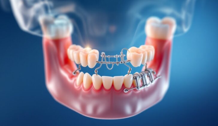
Non-surgical management, also known as closed reduction, involves stabilizing the fracture using a technique called maxillomandibular fixation (MMF). This method comes with several benefits, including lower costs as it doesn’t require surgery or a hospital stay, it being less invasive, and it doesn’t need a highly experienced doctor. However, we should not use MMF for patients who can’t follow instructions, have severe lung problems, have intellectual disabilities, are pregnant, or have multiple injuries.
Closed reduction is useful for fractures that are not displaced, which means the bones are still nicely lined up. It’s also suitable for developing, or when healthy teeth can provide stable closure of the mouth, or for patients who still have a good face shape and can open their mouth adequately.
In this procedure, doctors place Ivy loops, which are made from 24-gauge wire, between two stable teeth, followed by a smaller gauge wire that allows the Ivy loops to provide MMF. Arch bars with 24- and 26-gauge wires are also often used, which give extra stability and help stop the bone from moving while it heals. If the patient doesn’t have teeth we can attach dentures to the jaw using wires. Another option is to use MMF screws to fix the dentures.
On the other hand, surgical management, known as open reduction and internal rigid fixation (IRF), allows doctors to see the fracture site clearly. This method lets patients recover their normal mouth closure and speaking and eating capabilities quickly, and maintain their gum health. However, it does carry an increased risk of complications. The surgery could be done either from inside the mouth (intraoral) or from outside the mouth (extraoral), depending on the type and location of the fracture.
After surgery, doctors will prescribe painkillers, and antibiotics that target gram-positive bacteria. It’s important to keep a pair of wire cutters by the patient’s bedside in case they vomit. Generally, fixation (using wires or other materials to hold the bone in place) is kept for 4 to 6 weeks in most adult cases. However, if the fracture is minimal and located in an area with teeth, two weeks may be enough.
What else can Mandible Body Fracture be?
When treating a broken lower jaw, it is critical to thoroughly assess and monitor the patient’s overall health. The impact that causes such fractures could also potentially harm other parts of the body. Some possible conditions that may arise from this trauma include:
- Clogging of a major blood vessel in the neck due to blood clot formation
- Fracture at the base of the skull
- Presence of air pockets under the skin of the neck
- Accumulation of air or gas in the chest cavity which can affect the lungs
- Air surrounding the organs in the center of the chest cavity
- Injury to the spleen, often in the form of cuts or tears
Therefore, before deciding to operate on a lower jaw break, it’s essential that doctors identify and manage all associated injury or issues.
What to expect with Mandible Body Fracture
Procedures to correct fractures in the mandible, or the jawbone, whether through non-surgical (closed) or surgical (open) methods often result in successful bone healing. It’s also key to address any dental injuries at the same time as the fracture. This is because broken teeth can lead to infections that may disrupt the healing of the bone and might need to be removed.
Preserving the mandibular canines, the ‘pointy’ teeth in the jaw, is recommended if possible because they play an essential role in bite alignment. It’s important to note that managing fractures in individuals who don’t have teeth (edentulous) can be more challenging, often due to their older age and the presence of other health conditions.
Possible Complications When Diagnosed with Mandible Body Fracture
The main complication after a fracture, including mandibular (jaw) fractures, is often infection. This may result in improper healing of the bone (malunion or non-union) or a bone infection (osteomyelitis). Also, several factors can contribute to these complications:
- Oral infections
- Presence of teeth in the fracture line
- Alcohol abuse and chronic disease
- Delay before receiving treatment
- Poor patient cooperation
- Movement of fracture pieces
- Fracture of the plate
In addition, other complications can occur, such as delayed healing of the fractured jaw. If there is a bilateral fracture of the body of the jaw, along with fractures near the center of the jaw (parasymphyseal) or at the joints at the back of the jaw (condylar fractures), this can cause problems with breathing. The muscles may pull the part of the jaw behind the fracture backwards, causing the tongue to block the throat. Nerve damage can also occur and may include conditions like neuropraxia, where function could take 4 to 6 weeks to return, or neurotmesis, where function might take as long as 18 months to return.
Preventing Mandible Body Fracture
A broken jaw can make it difficult for patients to eat properly, which could lead to weight loss and poor nutrition. Not getting enough nutrients can cause the person to feel weak and hinder their recovery. As such, it’s important for patients to know what and how to eat after their operation. For the first three weeks post-surgery, they should stick to a full liquid diet, which can include high-protein drinks, pureed food, juices, and soups. They should also try to eat smaller portions more frequently to ensure they’re getting enough calories. In addition, patients are advised against smoking.
Poor nutrition can increase the risk of infections and slow down the healing of wounds, making it crucial for patients to monitor their weight on a weekly basis. This can help guide the necessary dietary adjustments to keep the body healthy. For patients who cannot read, pamphlets and simple picture booklets can be used to teach them about proper nutrition and maintaining a healthy weight while recovering from a broken jaw.


