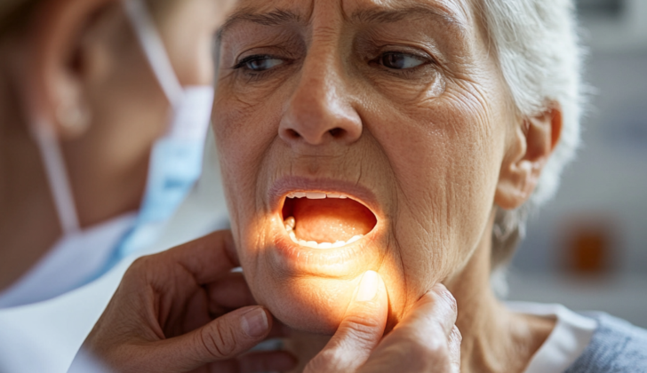
What is Sialolithiasis?
Sialolithiasis is a non-threatening condition where stones form within the ducts of the major saliva-producing glands in the mouth: parotid, submandibular, and sublingual glands. This condition is the primary cause of swelling in these glands, and it usually occurs between 1 in 10,000 to 1 in 30,000 people. Sometimes these stones can block the ducts, causing inflammation and possible infection known as sialadenitis. In rare cases, pus-filled pockets, or abscesses, can form. Symptoms often come and go and are tied to eating. They include swelling of the impacted gland and decreased saliva flow.
In cases where larger stones impact the tube that drains saliva from the floor of the mouth to the tongue (Wharton duct), doctors can often diagnose the condition through a physical check. With smaller stones, particularly in the tube carrying saliva from the cheek to the mouth (Stenson duct), diagnosis used to be made with traditional X-rays, sialography (an X-ray of the salivary gland), and digital subtraction sialography. Nowadays, doctors use more modern techniques like point-of-care ultrasound, CT scans, MRI scans, and sialoendoscopy, where they look directly into the glands with special equipment.
There are various treatment options available for sialolithiasis, which includes medication that stimulates saliva flow, massaging the stones out of the duct, and other procedures like interventional sialography, breaking up the stone (lithotripsy), sialoendoscopy, and surgery.
What Causes Sialolithiasis?
Researchers are still trying to fully understand what causes salivary stones, also known as salivary calculi. This is partly because the condition is quite rare, which makes large-scale studies difficult. There are two main factors that are believed to contribute to the formation of these stones: anatomical factors and compositional factors.
Anatomical factors are related to issues with the structure or function of your salivary glands. For instance, this could be a narrowing of the ducts that carry saliva (called duct stenosis) or inflammation. Compositional factors are related to the make-up of your saliva. For example, having too much calcium in your saliva or changes in how specific enzymes in your saliva function might contribute to stone formation.
Hard water doesn’t seem to increase the chances of getting salivary stones. Even in places with very hard water, there doesn’t seem to be an elevated risk. Studies also haven’t found a link between high levels of calcium in the body (hypercalcemia) and an increased risk of salivary stones. Other possibilities that are still being explored include whether not drinking enough fluids or side effects from medicines – like diuretics, which can decrease saliva production – could contribute to salivary stone formation.
Some recent discussion has suggested that tobacco smoking might be a risk factor. Smoking could potentially cause inflammation in the salivary ducts and decrease the production of an enzyme in your saliva called amylase, though this is still an area of ongoing research.
Risk Factors and Frequency for Sialolithiasis
Sialolithiasis is a condition where stones form in your salivary glands. It’s quite rare, affecting between 1 in 10,000 to 1 in 30,000 people. It’s most usually diagnosed when people are between 30 and 60 years old and is more common in men.
The most frequent place where these stones are found is in the submandibular gland, which is affected in about 85% of the cases. The reason for this is due to the way its duct is structured, with its opening located higher, leading to a stagnation of saliva. Another factor is that this gland produces a type of saliva that is more mucous and viscous than others, leading to an even slower flow of saliva.
Besides, the submandibular gland also produces more alkaline saliva, which makes it easier for the proteins and minerals in your saliva, like calcium and phosphate, to harden and form stones.
- About 15% of salivary gland stones occur in the parotid gland.
- Less than 5% happen within the sublingual and minor salivary glands.
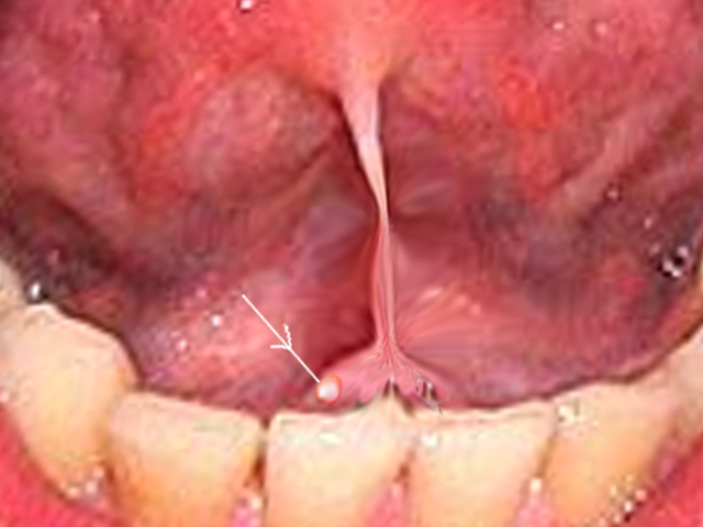
examination of the oral cavity.
Signs and Symptoms of Sialolithiasis
Sialolithiasis, a condition where stones form in the salivary glands, can be difficult to diagnose. This is because symptoms often only appear when the stones block the salivary ducts, causing swelling and pain, especially during meals. These symptoms usually occur only on one side of the face since only one salivary gland is typically affected.
A physical examination can reveal an uneven swelling of the gland, providing clues that the patient might have this condition. Stones can often be found in specific spots within the gland’s ducts, with 60% found in the parotid gland (near the ear) and 30% in the submandibular gland (under the jaw). If large enough, the stones might be visible when the doctor inspects the mouth, appearing white or yellow and round or oval in shape. If the stones are not visible, they can often be felt during a physical examination.
The size of salivary stones can vary significantly. Their diameter usually ranges from 2.1 to 10 mm, and only a small percentage (approximately 7.6%) grow larger than 15 mm. Typically, stones found in the submandibular gland are larger than those found in the parotid gland. They can also differ in weight, with most weighing around 300 mg, but they could weigh as little as 1 mg or as much as 5 g.
Testing for Sialolithiasis
In the past, traditional X-rays were used to initially diagnose salivary stones. These X-rays were good at spotting bigger, solid stones, but less effective for smaller and softer ones. Also, only about 80% of these stones are able to be seen by an X-ray, meaning a lot of them could go under the radar. While these conventional X-rays can still be helpful as an initial attempt towards diagnosis, using ultrasound or CT scans can offer a more certain diagnosis for these salivary stones and can also pinpoint their exact location.
Sialography is widely respected in the medical field as the most accurate way to diagnose salivary stones. In this technique, a contrast dye is injected into the salivary glands, making it possible to get a really clear picture of the size, position and appearance of the stones. We should keep in mind, however, that this technique does involve some exposure to radiation and some patients may have a reaction to the contrast dye used.
Non-contrast computed tomography (NCCT) is another commonly used medical imaging technique for salivary stones. It’s accurate when the stone is big enough, or the imaging is done with high detail. The perks are its high accuracy for detecting calcified (hard) stones, its quick image collection, and it’s widely available in medical facilities. The down sides include radiation exposure and it doesn’t provide a very detailed evaluation of the salivary ducts or any other underlying problems that may be causing salivary stones. Sometimes, a contrast dye is administered intravenously with NCCT to help visualize the stones better, but this is done cautiously as it could make tiny blood vessels appear to be calcifications, leading to a false diagnosis.
MRI sialography, which is similar to an NCCT in terms of accuracy, doesn’t need contrast dye and doesn’t involve radiation. Downsides here are the cost, the amount of time taken for image collection, and it’s not as widely available as NCCT or ultrasound technology.
Sialoendoscopy is a medical procedure that involves using a tiny camera to directly visualize the salivary stones and ducts. Its flawless accuracy, along with its increasing use in stone removal makes it a preferred method for diagnosis and treatment. It also has a lower chance of complications than conventional surgical techniques.
Ultrasonography is a non-invasive imaging technique for salivary stones. It usually shows the stones as bright spots on the images with shadows behind them. Its advantages include no radiation exposure, the possibility of seeing results in real-time, and it’s widely available. One thing to keep in mind is that it highly depends on the skill of the operator. It’s generally most effective for stones that are bigger than 2 to 3 mm in size.
Treatment Options for Sialolithiasis
When someone has sialolithiasis, which is the medical term for saliva stones, there are several treatment options that can be used. The first step usually involves conservative measures. This can involve actions like massaging around the area of the salivary gland, taking anti-inflammatory drugs, and using substances called sialogogues, which help to increase the amount of saliva that’s produced. If there are signs of infection, such as swollen lymph nodes in the neck, pus coming out of the saliva ducts, or redness and warmth around these ducts, antibiotics will usually be needed.
The next steps in treatment depend on how big the stones are, how many there are and where in the salivary gland they are located. If these initial treatments don’t work and the stone is less than 5mm in size, it’s in a submandibular gland (located under the chin) and it’s mobile, or can move around, then a procedure called endoscopy is usually tried first. This involves inserting a thin tube with a camera down your throat to look at and hopefully remove the stone. If other types of stones that are bigger or stuck are found, other surgical methods such as making a small cut in the mouth to access and remove the stone (transoral slitting) might be used.
Another strategy for dealing with some types of stones involves using External Shockwave Lithotripsy, or ESWL. This is a technique that uses sound waves to break the stone into smaller pieces that can hopefully be passed naturally or removed more easily. This technique is usually used when the stones are small, cannot be felt or seen under endoscopy. However, ESWL doesn’t tend to work well on larger stones.
Stones located within the parotid gland, which is in the cheeks, are normally removed using endoscopy if they’re under 7mm in size and can move around. If these stones can’t be managed successfully with endoscopy, then ESWL could be used as a next step, along with endoscopy to remove the broken-down stones. In a case where treatment with ESWL is not effective, a combined approach using a skin puncture and endoscopy might be used as long as the stone can still be seen under endoscopy.
It’s important to know that for both the under-the-chin salivary gland and the cheek salivary gland, surgical removal is usually considered a last resort. It may be needed if other treatments have failed, but because of the complexity of surgery and potential complications, doctors try to avoid it whenever possible.
What else can Sialolithiasis be?
Salivary gland swelling can be caused by several different conditions. When trying to identify the cause, physicians usually consider the following possibilities:
- Sialolithiasis (salivary stones)
- Sialadenitis (infection or inflammation of the salivary glands)
- Neoplasm (an abnormal growth, which could possibly be cancerous)
However, diagnosing the cause of salivary gland swelling can be complicated, because there are many conditions that affect the mouth and face and show similar symptoms. Because of this, doctors also pay close attention to what they find during a physical examination and what they learn from a patient’s health history. The following conditions can look much like salivary gland swelling:
- Cellulitis (a skin infection)
- Poor dentition and dental abscess formation (bad teeth and infections in the teeth or gums)
- Infection of the buccal or masticator space (infections in parts of the jaw or mouth)
- Herpes zoster (shingles)
- Neoplasm (again, an abnormal growth that can potentially be cancerous)
What to expect with Sialolithiasis
Sialolithiasis, a condition where stones form in the salivary glands, generally has a very good outcome. Most patients can be effectively treated with sialogogues (medicines that stimulate saliva production) and over-the-counter anti-inflammatory drugs. The minimally invasive procedures mentioned earlier are highly successful with very few complications compared to traditional surgical methods.
Moreover, sialadenectomy, a surgical procedure to remove a salivary gland, is rarely needed to treat sialolithiasis, thanks to modern treatment techniques.
Possible Complications When Diagnosed with Sialolithiasis
: Sialolithiasis, or salivary stones, can cause several complications. These include development of sialadenitis, an infection of the salivary gland, that could be either acute (short-term) or chronic (long-term). This happens when the stone obstructs the gland, preventing saliva from flowing and causing the gland to swell up and hurt.
The blocked salivary flow also means bacteria and debris can’t be flushed away from the salivary duct, which can lead to an infection. In cases where the obstruction persists for a long amount of time, it can harm the salivary gland’s acinar cells, creating local inflammation. If these conditions are not correctly treated, the gland may eventually become permanently fibrous and deteriorate.
The primary complications:
- Sialadenitis development (either acute or chronic)
- Pain and swelling of the salivary gland due to sialolith obstruction
- Bacterial infection due to blocked bacteria and debris removal
- Damage to the acinar cells in the salivary glands causing local inflammation
- Potential permanent fibrosis and decay of the gland if left untreated
Preventing Sialolithiasis
If you have been diagnosed with sialolithiasis, a condition where small stones form in your saliva glands, you should know that it usually has a very good outcome and can often be managed without aggressive treatment. The most common early signs of this condition are swelling of the gland and pain during meals, suggestive of the formation of a new stone. Normally, sialolithiasis happens without any clear reason. However, it can sometimes be caused by another issue, like the narrowing of your saliva duct (ductal stenosis) or a tumor.
It’s important that you stay aware of your symptoms and report back to your doctor if they get worse or keep coming back. This could be a sign that you need more detailed scans or that you need to see a specialist who treats conditions of the head and neck. Remember, open communication with your healthcare provider is key in managing and treating this condition effectively.


