What is Basal Cell Carcinoma?
Basal cell carcinoma (BCC), also formerly known as basal cell epithelioma, is the most common type of cancer in humans. BCC usually develops on skin that has been damaged by the sun, and it’s rare for it to appear on the moist tissues lining certain parts of the body, or on the palms of the hands and soles of the feet. Although this type of cancer grows slowly and rarely spreads to other areas of the body, it can cause severe damage and deformity to nearby tissues if not treated in time.
Upon examination, BCC typically shows up as small round bumps that are flesh-colored or pink, often with broken blood vessels on the surface. Although BCC can occur anywhere on the body, it most frequently appears on the head or neck. More than 26 different kinds of BCC have been recorded, including types like nodular, micronodular, superficial, morpheaform, infiltrative and fibroepithelial.
It’s crucial to note that most BCCs lack pigment, although some may contain varying amounts of melanin, a substance that gives color to the skin, eyes, and hair.
Regarding treatment, surgery is the most common approach and includes techniques such as excision (cutting out the tumor), electrodesiccation and curettage (burning and scraping away cancer cells), cryosurgery (using cold temperatures to kill cancer cells), and Mohs micrographic surgery (removing cancer in layers). These methods are typically used for localized BCC and have a high success rate, often curing over 95% of cases within five years.
What Causes Basal Cell Carcinoma?
The main cause of a type of skin cancer, known as basal cell carcinoma, is exposure to UV light. Both UVB and UVA light can play a role. Studies have shown that people who work outside are at a higher risk, especially in places further from the equator.
However, the amount of UV light and skin type aren’t the only factors to consider. Duration and intensity of the exposure, especially during childhood and adolescence, also contribute to the risk. Other factors that increase the risk of basal cell carcinoma include recreational sunlight exposure, using indoor tanning salons, UV light therapy, and occasional heavy sun exposure leading to sunburns. People with fair complexions, especially red-haired individuals, those who sunburn easily, and those who suffered blistering sunburns in childhood are at higher risk as well. Also, having a family history of basal cell carcinoma can put you at risk.
Despite the strong association with UV light exposure, about 20% of basal cell carcinomas occur on skin that isn’t exposed to the sun. They can also be caused by exposure to radiation, arsenic, immune system suppression, and genetic predisposition. Certain genetic syndromes, such as xeroderma pigmentosum, basal cell nevus syndrome (also known as Gorlin syndrome), Bazex–Dupre–Christol syndrome, and Rombo syndrome, can increase the risk of basal cell carcinoma.
There’s no evidence showing a connection with diet, but smoking appears to be a risk factor for women.
Risk Factors and Frequency for Basal Cell Carcinoma
BCC, or Basal Cell Carcinoma, is the most common type of skin cancer and its incidence rates are rising across the globe. It is more prevalent in men and tends to occur often in areas with a lot of UV exposure, such as places at extreme latitudes. The main predictor for developing BCC is a history of either BCC itself or squamous cell carcinoma (SCC), another type of skin cancer.
- People who have had BCC previously are at least ten times more likely to develop it again compared to those who have never had skin cancer.
- In the past 30 years, the number of BCC cases has increased by 20% to 80%.
- The likelihood of getting BCC increases with age, with the majority of people being diagnosed around the age of 68.
- Death from BCC is rare but it mainly happens in patients with weakened immune systems.
- The small percentage (1%) of BCC that does spread, or metastasize, typically affects the regional lymph nodes, bones, lungs, or skin.
- These aggressive tumour types include morpheaform, metatypical, basosquamous, and infiltrating patterns.
- The average age of death from BCC is higher than SCC, and the mortality rate is 0.12 per 100,000, adjusting for age.
- Risks of death increase with age, male gender – who get it at more than twice the rate of women, and persons of White race.
Signs and Symptoms of Basal Cell Carcinoma
Basal Cell Carcinoma (BCC) is a type of skin cancer that can appear in different forms. The three most common types are nodular, superficial, and morphea-like BCC.
Nodular BCC is usually a shiny, pink, or skin-colored lump on the skin with tiny visible blood vessels. This type of BCC can grow in size and even form an ulcer that makes the borders look raised or resemble a rodent ulcer. Nodular BCC often appears on the face, particularly the nose, cheeks, forehead, or near the nose and cheek fold, and the eyelids. Patients with nodular BCC often report a history of crusting and recurring bleeding, prompting them to seek medical advice. This type of BCC is also more common in individuals with darker skin.
- Presents as a shiny, pink- or flesh-colored lump
- Appear on the face
- May crust and bleed
Superficial BCCs are red, scaly spots or patches, which may contain tiny blood vessels. They usually appear on the shoulders, chest, or back, and there could be multiple lesions. Sometimes superficial BCC can look like skin conditions such as eczema or psoriasis. If you have a persistent red, scaly spot, it might be a superficial BCC. Over time, parts of superficial BCC can evolve into nodular BCC.
- Presents as red, scaly spots or patches
- Appear on shoulders, chest, or back
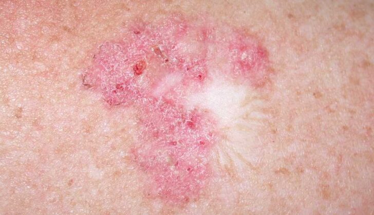
The morpheaform subtype of BCC typically looks like a white- or flesh-colored hard patch with unclear borders. It may resemble a scar or plaque of morphea. The surface of the lesion is usually smooth, although there can be crusts with underlying erosion or sores, and small lumps. Tiny visible blood vessels may also be present. This type of BCC generally behaves more aggressively and can cause extensive local tissue damage.
- Presents as white- or flesh-colored hard patch
- Surface typically smooth
- Can cause extensive local tissue damage

Testing for Basal Cell Carcinoma
If you’re suspected of having basal cell carcinoma (BCC), a type of skin cancer, your doctor will need to do a skin biopsy for confirmation. There are different types of biopsies: shave, punch, or excisional, and it’s important to sample a bit of the dermis layer of your skin as part of this. These techniques help to tell apart superficial BCC from other invasive types and are about 80% accurate.
Your healthcare provider will do a comprehensive skin examination as people who have one skin cancer often have other cancers or pre-cancers elsewhere on their skin. They are also at a higher risk for developing a more dangerous type of skin cancer called malignant melanoma. Their findings will be documented with photos or digital images. They may take samples from any suspicious areas for further testing, and in some cases, advanced imaging studies might be necessary. For instance, they might be concerned about the involvement of your parotid gland, muscles, deep soft tissues, eye socket, bones, or if the cancer has spread along your nerves. Patients with a history of BCC will need ongoing monitoring, potentially for their entire lives, especially if they’ve had multiple or high-risk tumors.
Dermoscopy, a skin imaging technique, can also help with the diagnosis. Under the dermascope, BCC often shows up with a distinct pattern of blood vessels. Other signs include multiple blue-gray spots, leaf-like structures, large blue-gray nests, and spoke-wheel areas. Unlike many pigmented lesions, there’s no pigment network in BCC.
Treatment Options for Basal Cell Carcinoma
The choice of treatment for Basal Cell Carcinoma (BCC) depends on different factors like the patient’s age and gender, size of the lesion, and the location and type of lesion. There isn’t one ideal treatment for all cases. To confirm a suspected BCC diagnosis, a biopsy is necessary. The main goals of treatment are to remove the tumor completely to prevent future reoccurrences, address any functional issues caused by the tumor, and achieve the best cosmetic outcomes as many BCCs happen on visible areas like the face.
Usually, surgical methods are used to treat BCC, but some types can be treated with medical treatment or radiation therapy as well. Different treatment options include Mohs micrographic surgery (MMS), standard surgical excision, EDC, radiation, photodynamic therapy, cryosurgery, topical treatments, and systemic medications such as Vismodegib. Recurrence rates for primary BCC using these treatments vary, Michael surgery has the lowest at 1%, while surgical excision has one of the highest at 10.1%.
Mohs surgery offers the best long-term results for treating BCC. It is considered the gold standard, especially for high-risk BCCs and recurrent BCCs, because of its high success rate and its ability to conserve healthy tissue. This success rate is due to the thorough examination of the tissue margins when compared with a standard vertical sectioning that only examines less than 1% of the outer peripheral and deep margins.
After standard surgical excision, a pathologic analysis is carried out. For well-defined tumors smaller than 2 centimeters, four-millimeter margins are typically sufficient. For facial lesions, a simple excision with narrow margins often doesn’t fully remove the tumor. EDC is often used for low-risk BCCs. While it’s one of the fastest and least expensive treatment methods, the downside is that it usually results in a white atrophic scar that can cause cosmetic issues.
Radiation therapy is an option for treating BCC in cases where surgery isn’t advisable. It can also be used as added treatment when further surgery could affect major nerves or other critical structures. Disadvantages of radiation include cost, potential negative cosmetic results, long treatment duration, and increased risk for future skin cancers. Notably, radiation therapy scars often get worse over time, unlike scars from surgery, which tend to improve.
Cryosurgery is another option for low-risk BCCs that involves applying liquid nitrogen to the visible tumor and a small border of normal skin. This procedure is straightforward, relatively quick, and a good fit for patients wishing to avoid invasive surgery. However, the treated area may be painful and swollen afterward, and there may be long-lasting changes in skin color and hypertrophic scarring.
Topical therapies are another BCC treatment option. The Food and Drug Administration (FDA) has approved two topical therapies, 5-fluorouracil (5-FU) and Imiquimod 5% cream, to treat superficial BCC. However, these therapies come with common side effects like redness, itchy skin, pain, swelling, changes in skin color, scabbing, bleeding, and skin erosion. Another disadvantage is that there is no way to confirm if the entire tumor has been cleared.
For cases of advanced or metastatic BCC that can’t be treated using conventional methods, a newer treatment involves Hedgehog pathway inhibitors. However, the side effects of these treatments can cause up to 55% of patients to stop treatment. These side effects may include nausea, changes in taste, muscle spasms, hair loss, weight loss, diarrhea, and fatigue.
In some cases, such as with elderly patients, those with weakened immune systems, and patients whose BCCs are in cosmetically sensitive areas, nonsurgical treatments may be preferred.
What else can Basal Cell Carcinoma be?
When diagnosing Basal Cell Carcinoma (BCC), which is a type of skin cancer, doctors consider other conditions that may show similar skin changes. These include skin tumors that affect hair follicles or sweat and oil glands, as well as a different type of skin cancer known as Squamous Cell Carcinoma (SCC).
There are different forms of BCC that can be mistaken for other conditions. For example, a BCC variant known as nodular BCC can be mistaken for a trichoblastoma or trichoepithelioma, both of which are types of benign skin tumors. Superficial BCC, on the other hand, can look similar to inflammatory skin conditions such as psoriasis and eczema. There’s also a type of BCC known as morphea-like BCC that can look like a morphea plaque, a condition that causes thickened skin, or a scar.
In these situations, an examination of tissue under a microscope, also known as a histopathological examination, can help confirm the diagnosis of BCC.
What to expect with Basal Cell Carcinoma
Basal cell carcinoma (BCC) rarely leads to fatal results. Instead, the main concern with BCC lies in its potential to recur, or return, after initial treatment. This risk of recurrence largely depends on where the BCC is located and what its clinical and histopathological (cellular structure and appearance) features are.
With respect to location, the trunk and limbs are considered low risk; the forehead, cheeks, chin, neck, and scalp bear intermediate risk, while the areas in the middle of the face, the nose, ears, and around openings in the body, or on embryonic fusion planes, are designated as high-risk areas.
The clinical and histopathological features that influence the risk of recurrence include the size of the BCC, the type of cells involved, and whether the tumor is a first-time occurrence or a recurrence.
As for the prognosis, or the likely course of the disease, it varies depending on these factors. A primary superficial BCC or a primary nodular BCC smaller than 1cm in an intermediate-risk location, or smaller than 2cm in a low-risk area, generally has a good prognosis.
However, a recurrent superficial BCC, nodular BCC smaller than 1cm in high-risk location or smaller than 2cm in an intermediate-risk area or larger than 2cm in a low-risk location, typically has an intermediate prognosis.
The prognosis is poor for larger nodular BCC (larger than 1cm) in a high-risk location, aggressive or infiltrating types of BCC, morpheaform BCC (which has a sclerotic, or hardening, feature), or any recurrent tumor apart from superficial BCC, all because these carry an extremely high risk of recurrence.


