What is Cutaneous Melanoacanthoma?
Melanoacanthoma is an uncommon, harmless skin growth. It is generally seen in older adults around 65 years of age, and more frequently in men. The exact cause of this skin growth is unknown. Some suggest it might be a version of a skin condition with too much pigment, known as seborrheic keratosis. Melanoacanthoma usually appears as a solitary, slow-growing black bump on areas like the head, neck, trunk, or limbs.
Diagnosing melanoacanthoma can be tricky as it might look like a more serious skin condition under certain skin examination methods. Hence, taking a sample (biopsy) and examining it under a microscope is crucial. During such an analysis, the skin sample shows certain specific features, such as increased skin thickness, skin-cell growth, and wart-like growth. Certain additional tests may be required to confirm the diagnosis.
The primary treatment for melanoacanthoma is the complete removal of the growth. This is necessary because, if not completely removed, the growth may persist, continue to grow or reappear. Patients who have had a melanoacanthoma should be monitored for any new or returning growths.
Melanoacanthoma can also appear in the mouth or other soft tissue areas. This version, known as oral melanoacanthoma, is believed to be a reaction, rather than a tumorous growth. There’s some debate over whether seborrheic keratosis and melanoacanthoma are separate conditions, but they are currently identified as distinct due to their unique characterstics and treatment needs.
Melanoacanthoma has also been linked to some other conditions. For example, successive cases of oral melanoacanthoma have been seen in patients with Laugier-Hunziker syndrome. Also, a case of melanoacanthoma on the lower lip was associated with two conditions: plasma cell cheilitis and increased growth of a type of immune cell in the lower lip.
What Causes Cutaneous Melanoacanthoma?
The exact cause of melanoacanthoma, a type of skin condition, is currently unknown. However, it’s thought that it could be a reaction to something. This idea comes from the observation that a type of cells called dendritic melanocytes often appear in skin conditions that are known to be reactive, like lichen simplex chronicus, warts, and squamous cell carcinoma.
Dendritic melanocytes can be found throughout all layers of the skin in a melanoacanthoma. These cells usually appear in areas of the skin that have been injured, which has led to the idea that they can cause irritation, lead to the formation of skin lesions, and make these lesions grow quickly.
There are also certain superficial changes that can take place in the skin, such as the formation of ulcers. This can happen due to poor blood flow, rubbing, pressure when lying down, or further injuries to the skin.
It’s worth noting that even though melanoacanthoma shares some characteristics with other reactive skin conditions, it is a type of skin growth or neoplasm. This is different from a similar condition, oral melanoacanthoma, which is thought to be purely a reactive condition. Because of these differences, some aspects of what causes melanoacanthoma are still unknown.
Risk Factors and Frequency for Cutaneous Melanoacanthoma
Melanoacanthoma is a condition whose exact occurrence rates are still not clear due to inconclusive research results. Some studies indicate that it appears in 1 of every 100,000 pathology tests, while others suggest it may be present in around 1% to 38% of related skin issues like seborrheic keratoses. The variability in the studies stems from differences in including pigmented seborrheic keratosis, an issue that may have been overlooked when diagnosing melanoacanthoma in the past.
This condition generally affects older adults; typically those above 60 years of age, with the average age being 65. Nevertheless, it can occur in adults of all ages. Although most reported cases involve men, there doesn’t seem to be a significant gender pattern. Melanoacanthoma is a worldwide condition, with reports of cases from various countries.
- Out of 52 cases studied, 28 were men.
- The condition has been reported in countries including Belgium, Brazil, Canada, India, Italy, Japan, South Korea, Spain, and the USA.
The race or nationality of 43 patients with melanoacanthoma has been identified, with the most cases (42%) reported in white patients. Additionally, the condition has been found in patients from various ethnic backgrounds, including Hispanic, Indian, Asian, Black, Brazilian, Haitian-Creole, and Italian.
Signs and Symptoms of Cutaneous Melanoacanthoma
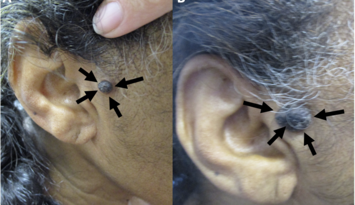
Melanoacanthoma is a condition that usually presents itself as a pigmented growth on the skin that is typically harmless and grows slowly. It can appear as a small bump, larger nodule, or a patch of skin. Despite being rare, they can occasionally grow quickly, cause itchiness or discomfort, or become irritated. People might have melanoacanthomas for several weeks or even years before they are diagnosed. Their size can vary greatly, ranging from 0.2 cm to 15 cm, with an average size of about 2 cm.
Interestingly, about half of these growths are black, but they can also show a combination of colors such as black and blue, black and gray, or black and brown, which can make them look similar to a type of skin cancer called melanoma. Sometimes, the surface of a melanoacanthoma can have a lumpy or warty appearance.
Typically, melanoacanthomas appear on the head, neck, or trunk. However, they have been occasionally reported on the arms and legs, genitals, or buttocks. It’s rare to see multiple lesions at once, which sets it apart from a similar condition known as oral melanoacanthomas. In very rare cases, a melanoacanthoma might present as a cutaneous horn – a protrusion from the skin – or grow outward like a wart or a squamous cell carcinoma – a type of skin cancer.
Testing for Cutaneous Melanoacanthoma
To determine if someone has melanoacanthoma, a skin condition, a small sample or biopsy of the skin lesion is typically taken. This sample gives the pathologist enough tissue to examine under a microscope, which will hopefully allow them to give an accurate diagnosis. Two other promising diagnostic tools are dermoscopy and reflectance confocal microscopy. However, these techniques alone are not enough to diagnose melanoacanthoma based on the current scientific knowledge.
Dermoscopy is a technique that uses a special magnifying lens to examine skin lesions. The appearance of melanoacanthoma under the dermoscope can vary. Sometimes, it can look like problematic skin conditions, including melanoma, a type of skin cancer. Other times, it can look like simple, harmless skin changes. Therefore, it’s important to interpret these findings alongside other tests and examinations.
Reflectance confocal microscopy is another tool that provides detailed images of the skin layers. When used to study melanoacanthoma, it showed specific features such as surface depressions filled with skin debris, a honeycomb pattern in the spinous-granular skin layer, and the presence of specific cells like dendritic cells and melanophages in the dermal layer. This tool gives a lot of detailed data, but it doesn’t make the final diagnosis. Instead, the results need to be interpreted in the context of the whole picture, including the patient’s symptoms and other diagnostic tests.
Treatment Options for Cutaneous Melanoacanthoma
Often mistaken for a type of skin cancer called melanoma upon initial examination, melanoacanthomas are, generally, surgically removed during biopsy or through a simple excision process. However, there have been instances where a partial removal during a biopsy resulted in the continued presence of the lesion, particularly noticed before the ear in one patient. In another reported case, a nose melanoacanthoma came back six months after it was removed.
Aside from surgical removal, there are other treatment options for melanoacanthomas. For example, doctors may opt to use cryotherapy, a method that involves using liquid nitrogen to freeze and remove the growths. Topical 5-fluorouracil 5% cream, a type of chemotherapy drug often used in a cream form to treat certain skin conditions, can also be considered in treatment plans.
What else can Cutaneous Melanoacanthoma be?
Even though melanoacanthomas are harmless skin conditions, it’s important to rule out other similar conditions such as:
- Actinic keratosis: A type of skin damage caused by the sun. Under microscope examination, it will show abnormal cells.
- Basal cell carcinoma: A type of skin cancer that may look like melanoacanthoma. A close examination of its tissue composition will help identify it.
- Melanocytic nevus: Also known as a mole, if it’s raised, it can be hard to tell apart from a melanoacanthoma without examining the tissue closely. Some are flat and can be visually differentiated from a melanoacanthoma.
- Melanoma: A serious type of skin cancer that may closely resemble a melanoacanthoma. Special stain techniques may be required to accurately identify it under microscope examination.
- Pigmented spindle cell (Spitz) nevus: Another type of skin mole.
Also on the list are:
- Seborrheic keratosis: A benign skin growth where the pigment and pigmented cells are mostly found in the lower skin layers. However, in the case of a melanoacanthoma, the pigment is from certain cells that are spread across all layers of the skin.
- Solar lentigo: Also known as sun spots. These are generally flat, while a melanoacanthoma is usually raised.
- Squamous cell carcinoma: A major type of skin cancer that can be ruled out by the lack of unusual cells and a high number of cell divisions under microscope examination.
- Verruca vulgaris: Commonly refers to skin warts. Their absence is ruled out by lack of certain cell changes.
All of these conditions may closely resemble a melanoacanthoma, making it essential for a comprehensive diagnosis to clearly distinguish between them.
What to expect with Cutaneous Melanoacanthoma
The outlook for someone with melanoacanthoma is generally very good. This condition is non-cancerous and doesn’t lead to any body-wide health issues. After the lesion is removed, it usually doesn’t come back, although some scarring or other signs of the removal may still be there. In rare cases, the lesions may still be present or come back, but they are very unlikely to turn into cancer.
Possible Complications When Diagnosed with Cutaneous Melanoacanthoma
Melanoacanthoma is a harmless skin growth that doesn’t transform into a cancerous lesion. Yet, one should be careful as it might appear near a harmful lesion, which can make diagnosis more difficult. These lesions usually grow slowly, but occasionally, they can grow rapidly or even exceed 10 cm in size, becoming quite large.
Despite their benign nature, both small and large lesions can become irritated and cause discomfort. This might happen due to constant friction with the neighboring skin or even clothing. In rare cases, larger lesions can open up or develop a superficial infection.
Preventing Cutaneous Melanoacanthoma
Simply put, melanoacanthomas are harmless skin anomalies that can look like other benign (non-cancerous) or malignant (cancerous) skin conditions such as seborrheic keratosis or melanoma. Hence, if older people spot skin lesions that look like melanoacanthomas, they should get them checked out.
Non-invasive methods such as clinical observation, dermoscopy (skin surface microscopy), and reflectance confocal microscopy (a type of skin imaging) can be used for examination, but these alone can’t provide a definite diagnosis. So, if there’s a chance that the skin lesion might be a melanoacanthoma, doctors should take a biopsy (a sample of tissue for examination) of the lesion to make sure of the diagnosis and rule out other conditions.


