What is Dermatomyositis?
Dermatomyositis is a rare condition where the immune system mistakenly attacks the muscles, causing weakness and a skin rash. It’s part of a group of muscle diseases referred to as idiopathic inflammatory myopathies. Each of these conditions cause muscle weakness, but they can affect different muscle groups and show different results in tissue examinations. Dermatomyositis is recognized by distinct skin symptoms and typically impacts the muscles closer to the center of the body. Nevertheless, it can also affect other parts of the body, such as the lungs, heart, and digestive system.
A remarkable point to consider is that a significant number of patients with dermatomyositis might have cancer, which can impact how the condition progresses. While most people with dermatomyositis experience muscle weakness and skin symptoms, there are also different forms of the condition.
One variant is Clinically Amyopathic Dermatomyositis (CADM), where patients have the typical skin symptoms of dermatomyositis, but don’t experience muscle weakness. This subtype is then further divided into hypomyopathic and amyopathic dermatomyositis. People with hypomyopathic dermatomyositis don’t exhibit muscle weakness, however, tests can indicate inflammation in their muscles. On the other hand, patients with amyopathic dermatomyositis show no signs of muscle weakness, and tests also don’t provide any evidence of muscle inflammation.
What Causes Dermatomyositis?
While the exact cause of dermatomyositis is unknown, it is believed that a combination of genetic, immune system, and environmental factors may play a part in this condition.
From a genetic perspective, research shows that people with certain types of a gene called the human leukocyte antigen (HLA) are more likely to get dermatomyositis. This includes types such as HLA-A*68 seen in North American whites, HLA-DRB1*0301 in African Americans, and HLA-DQA1*0104 and HLA-DRB1*07 in the Han Chinese population.
Next, there’s the immune system factor. Some people with dermatomyositis have been found to have autoantibodies – antibodies that mistakenly attack one’s own cells. However, it’s still unclear whether these autoantibodies are a cause or an effect of the disease.
Lastly, the environment also appears to have an influence. Viruses like the Coxsackie B virus, enterovirus, or parvovirus have been suspected of triggering dermatomyositis. The exact way in which these viruses could lead to the disease is still under study, but it could involve actions such as changing cell proteins, disrupting the body’s self-tolerance, or activating B cells, which are a type of white blood cell.
Also, certain medications, including cancer drugs like hydroxyurea and cyclophosphamide, antibiotics like penicillin, and non-steroidal anti-inflammatory drugs, may provoke dermatomyositis. Even some vaccines have been linked to the condition.
As for radiation, it has been observed that dermatomyositis occurs more often in women who have been exposed to a high intensity of ultraviolet rays.
Risk Factors and Frequency for Dermatomyositis
Dermatomyositis is a rare disease that affects fewer than 10 people per million every year, as identified in a study done in Minnesota, USA. Out of all those suffering, about 21% have a specific type called amyopathic dermatomyositis. The disease typically occurs in people aged between 40 and 50, with the usual age of diagnosis being around 44 years old. It’s prevalent more among women than men. Also, the occurrence differs in various places. Southern Europe has more cases compared to Northern Europe, and there’s also a higher rate of the disease in cities than rural areas in Quebec, Canada. There is a suggestion from a study in Pennsylvania that poor air quality may cause more cases of one type of the disease, clinically amyopathic dermatomyositis.
- Dermatomyositis, a rare disease, affects around 9.63 per 1,000,000 people.
- About 21% of all cases are of the amyopathic subtype.
- This disease typically shows up in people between the ages of 40 and 50.
- The average age at diagnosis is about 44 years old.
- Dermatomyositis occurs more often in women than in men.
- The rates of this disease are higher in Southern Europe compared to Northern Europe.
- In Quebec, it has been found that the disease is more common in urban areas.
- A study in Pennsylvania showed a connection between high airborne pollution areas and increased cases of a type of the disease called clinically amyopathic dermatomyositis.
Signs and Symptoms of Dermatomyositis
When doctors suspect a person may be suffering from dermatomyositis, they typically carry out a comprehensive examination. The aim is to look for telltale signs and symptoms of the disease, rule out other possible causes of muscle weakness, see if other parts of the body are affected, such as the lungs, heart, or esophagus, and look for any signs a person might have cancer.
The most common symptoms of dermatomyositis are muscle weakness and skin problems. The rate at which the disease develops can vary – it may come on slowly or suddenly, with periods where symptoms get better or worse.
- Muscle weakness: Most patients complain about gradually deteriorating muscle strength which may make it hard for them to do simple tasks like climbing stairs, standing up from a chair, lifting things, grooming, and even lifting their head while lying down. While muscle pain and stiffness are uncommon, they can occur. If the disease is severe, patients could also experience difficulty swallowing or speaking. During the physical exam, the doctor may note weaker muscles around the shoulders, hips, and neck. Typically, the patient does not experience significant tenderness, and the strength in their hands and feet remains unchanged. Reflexes and muscle size are usually normal unless the disease is severe and has been present for a long time.
- Skin Changes: Skin changes can occur before or at the same time as muscle weakness. Patients can develop varying types of rashes, be sensitive to sunlight, have itchy skin, or see changes in their skin color. The disease can also affect nails and cause hair loss. The doctor will look for:
- Gottron papules: Small, discolored, possibly scaly or ulcerated, bumps over the finger joints.
- Heliotrope rash: A purplish or reddish rash over the upper eyelids, which could be accompanied by swelling. This sign might not be evident, especially in individuals with darker skin tones.
- Gottron sign: Red patches over the elbows or knees.
- Facial erythema: Redness over the cheeks and the bridge of the nose, including the areas around the nose and mouth. The rash may cover the forehead and extend to the ears as well.
- Shawl sign: Redness over the back of the neck, upper back, and shoulders, and sometimes, the upper arms.
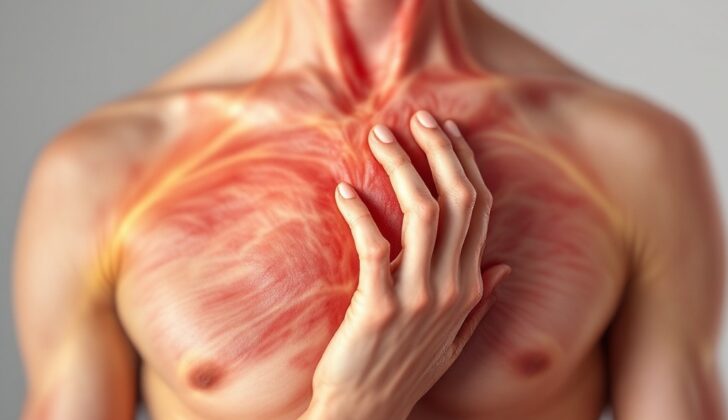
- V sign: Red patches on the front of the neck and upper chest.
- Poikiloderma: Skin becoming thinner with changes in pigmentation and the formation of small, dilated blood vessels. These changes can occur in areas exposed to sunlight and areas that aren’t.
- Holster sign: Poikiloderma present on the sides of the thighs.
- Changes to the skin around the nails: Presence of small, dilated blood vessels and overgrowth of the cuticle.
- Mechanic’s hands: Thickened, cracked skin on the palms and sides of fingers.
- Scalp involvement: Diffuse poikiloderma, with scaling and itching.
- Calcinosis cutis: Calcium deposits in the skin.
- Joints: Dermatomyositis can cause painful or swollen joints, particularly in the hands.
- Respiratory symptoms: Patients might be short of breath, find it difficult to exercise, or have a dry cough due to an underlying condition known as interstitial lung disease. You may notice reduced chest movement due to weakness in the respiratory muscles.
- Esophageal symptoms: Due to muscle weakness, patients might find it difficult to swallow both solids and liquids. They might also experience acid reflux.
- Other findings: Some patients might also experience Raynaud’s phenomenon (changes in color of fingers and toes in response to changes in temperature or emotional stress), stomach ulcers, heart symptoms, fever, fatigue, and weight loss. If a patient is male, older, experiencing difficulty swallowing, and does not have interstitial lung disease, this may point to an increased likelihood of an underlying cancer.
In addition to the physical exam and history, the doctor might ask about any medication the patient is taking and will ask about the family’s health history. Patients should also undergo cancer screening exams appropriate for their age, such as breast exams, pelvic exams, testicular exams, and rectal exams.
Testing for Dermatomyositis
If you have suspected dermatomyositis, your doctor may order initial tests on muscle enzymes such as creatine kinase, aldolase, lactate dehydrogenase, and some types of aminotransferase. These tests can help to guide further tests and evaluate how your body is responding to treatment. Before muscle weakness appears, you may see a rise in muscle enzymes.
While Antinuclear antibodies can be found in most patients with dermatomyositis, they are not usually used for a diagnosis. Instead, testing focuses on myositis specific autoantibodies, which are found in about 30% of patients. These antibodies can provide useful information on predicting how the disease will progress and what organs might be involved. Usually, Anti-Jo is the most common form of this antibody found in dermatomyositis.
Electromyography is used to identify the muscle groups that are most affected by the disease, guiding doctors on where to seek biopsy samples. It also helps distinguish dermatomyositis from other conditions that affect the nerves.
Every dermatomyositis patient should also have a chest X-ray to screen for lung disease. If you have respiratory symptoms or the X-ray isn’t clear, you may need computer tomography of your chest and further lung tests. MRI imaging of skeletal muscles is a good way to evaluate inflammation caused by dermatomyositis.
Doctors confirm the diagnosis of dermatomyositis using muscle biopsy. The biopsy can exclude other causes of muscle weakness or skin rash. Choosing the right muscle for biopsy is crucial; it should be done on weak muscles or those identified as abnormal by electromyography.
Typical tests may include full blood count, liver function tests, and inflammatory markers. Your doctor may also test your thyroid hormones to rule out hypothyroidism and get an ECG to look for hidden abnormalities in the heart. Lung function tests may be done to assess the severity of any pulmonary involvement.
During the first five years after a diagnosis, dermatomyositis patients are at high risk of having an underlying cancer. An initial cancer screening should take place at diagnosis, which are usually appropriate to the patient’s age and sex. The screening could range from blood tests, imaging modalities, to even biopsies.
There are several classification criteria historically used for dermatomyositis. These include the Bohan and Peter classification, the Targoff criteria and the Dalakas and Hohlfeld’s criteria. The most recent classification system was released by the European League Against Rheumatism and the American College of Rheumatology in 2017. This classification solves several limitations of the previous ones and contains clinical, lab, and muscle biopsy criteria to calculate the probability of having the disease.
Treatment Options for Dermatomyositis
The main goals of treating dermatomyositis, a condition that causes muscle weakness and skin problems, are to control muscle weakness, handle any skin issues and manage any other underlying problems.
The initial treatment for muscle weakness in dermatomyositis typically involves high-dose prednisolone, a type of steroid. The aim is to improve muscle strength and decrease muscle enzyme levels. Patients are usually given high doses of this drug for a few months and monitored for improvements. After about six weeks, muscle enzyme levels should be back to normal, and after about three months, muscle weakness should start to get better. Once doctors note improvement, they gradually reduce the dosage of prednisolone over time, usually over a course of nine to twelve months.
If the initial treatment doesn’t work, doctors may explore other possible causes of muscle weakness, such as hormone imbalances, other types of muscle diseases, or undiagnosed cancer, and consider adding immune system-suppressing drugs to help.
These drugs can help manage the side effects of long-term use of steroids, such as bone thinning, increased risk of infections, changes in appearance, and an increased chance of developing diabetes. They’re typically added in cases of very severe muscle disease, ongoing health conditions like diabetes, or serious complications like lung disease or problems with the esophagus.
For those whose conditions don’t improve with this combined treatment, other drugs like rituximab, mycophenolate mofetil, or intravenous immunoglobulin may be recommended. It’s important to monitor patients regularly for any side effects from these drugs.
For the skin disease aspect of dermatomyositis, methods to manage the problem can include avoidance of sunlight, using sun-protective clothing and sunscreen, and medication. Itching may be managed with local ointments and oral medication. For systemic treatment, hydroxychloroquine and methotrexate are commonly used.
Calcinosis, a condition where calcium builds up in the skin, is more common in children with dermatomyositis and can be managed with drugs like diltiazem that block the channels where calcium enters cells. In some cases, removing the buildups surgically might be needed.
Physical therapy and rehabilitation offer significant benefits for patients with dermatomyositis. Active exercise programs and range of motion exercises can help prevent the shortening of muscles or tendons. For patients with difficulty swallowing, working with a speech therapist might be useful, and they may need to take precautions to prevent choking.
Patients taking long-term steroids might require therapy to prevent bone thinning, and those on high-dose steroids or immune-suppressing medications should be considered for preventative treatment against a specific type of pneumonia. Also, all patients should receive the appropriate vaccinations before starting immune-suppressing drugs.
What else can Dermatomyositis be?
Just like dermatomyositis, several other conditions can also cause muscle weakness. These need to be ruled out by carrying out a health history, physical examination, and further tests.
- Inclusion body myositis: It’s different from dermatomyositis as it causes uneven muscle weakness and affects muscles like the wrist and finger flexors. There might be noticeable muscle loss, unlike dermatomyositis. Specific muscle biopsy results will highlight this disease. Treatment usually doesn’t respond to corticosteroids.
- Drug-induced myopathy: Certain medication can lead to muscle discomfort due to drug reactions. Common drugs that cause this include statins, alcohol, colchicine, glucocorticoids, and other medicines. This condition might have no skin-related symptoms and can be identified through muscle biopsy.
- Hypothyroidism: This and dermatomyositis can both cause muscle weakness and increased muscle breakdown. Other symptoms of hypothyroidism and results from muscle biopsy and thyroid tests can help to differentiate between the two.
- Myasthenia gravis: This condition mainly weakens the eye and throat muscles, unlike dermatomyositis. It is also linked with specific antibodies and doesn’t lead to increased muscle breakdown.
- Polymyalgia rheumatica: This mainly causes muscle pain and stiffness around the shoulder and hip areas. It can be differentiated from dermatomyositis by the presence of inflammation, absence of increased muscle breakdown, and normal muscle strength. It mostly affects people over the age of 50.
Further conditions that should be considered include muscular dystrophy, motor neuron diseases, neuropathy, inherited metabolic disorders, and myasthenia gravis.
Diagnosing dermatomyositis is often based on specific skin symptoms along with muscle weakness. A procedure called electromyography helps to distinguish between a weakness caused by nerve damage or muscle disease. Muscle biopsy can also be helpful to rule out other conditions.
What to expect with Dermatomyositis
The death rate for dermatomyositis, a skin and muscle disease, is thought to be about 10%. In the first year of the illness, the death rate is particularly high. The deadliest complications of dermatomyositis are cancer, lung complications, and heart disease due to blocked blood vessels. Certain factors suggest a high likelihood of death and a poor prognosis:
* Being older in age
* Starting treatment more than six months after symptoms begin
* Experiencing severe muscle weakness at the first doctor’s visit
* Having difficulty swallowing
* Affected lungs, either with weak respiratory muscles or a condition called interstitial lung disease
* Heart complications
* The presence of cancer
Of the patients who survive, 65% regain normal strength, 34% suffer mild disability, and only 16% experience no disability. With appropriate treatment, 20% of patients achieve complete recovery or ‘remission’, while 80% cope with a long-lasting or recurrent condition.
Possible Complications When Diagnosed with Dermatomyositis
Respiratory diseases are one of the complications of dermatomyositis. This can involve difficulty breathing, aspiration pneumonia, or a disease that affects areas of the lungs. This last condition known as interstitial lung disease (ILD), is common in about a third of patients with dermatomyositis. Aspiration pneumonia, which is a lung infection caused by inhaling food, drink, or saliva into the lungs, can happen because of weakness in the respiratory muscles and can lead to serious health problems or even death.
Another risk for patients with dermatomyositis is developing cancer. This happens in about 24% of cases. Some factors that increase this risk include being older, not having interstitial lung disease, severe skin conditions, presence of certain antibodies, lack of specific antibodies, resistance to treatment, and a history of previous cancer with a return of the disease. The most common types of cancers seen are of the ovary, lung, pancreas, stomach, and colon, as well as a type of cancer called non-Hodgkin lymphoma. The risk of cancer is at its highest in the first year of the disease but remains high for up to five years. Even after this period, the risk of developing cancer remains higher than for people without the disease.
Heart disease is another potential problem for people with dermatomyositis. Even though it often doesn’t show any symptoms, an EKG can sometimes detect irregular heart rhythms and other conductive problems. The heart might be affected directly, leading to inflammation (myocarditis), congestive heart failure, or coronary artery disease.
Weak muscles in the throat and esophagus due to dermatomyositis can cause difficulty swallowing, which could potentially lead to malnutrition and increase the risk of inhaling food or drink into the lungs.
Additional complications could include calcinosis (the formation of calcium deposits in the skin), muscle wasting, and joint contractures (permanent shortening of a muscle or joint).
Preventing Dermatomyositis
Care must be taken to educate both patients and their families about the nature of the disease, how it advances, what the potential outcome is, and the need to keep an eye out for any negative effects from the medication they’re taking. Here are additional recommendations:
Try and prevent sun exposure where possible – it’s crucial for patients to steer clear of sunlight, regularly apply sunscreen, and adopt other precautions such as donning wide-brimmed hats and clothing that fully covers the body. This can help ensure that the photosensitive skin rash characteristic of the disease doesn’t get any worse.
If a patient has severe muscle inflammation, they might need to follow a high-protein diet. If they also have esophageal dysfunction, they may need to follow a specific diet and make sure to prop up the head of their bed following meals.
Physical exercise and rehabilitation is a vital aspect of managing the disease. Activities like aerobic exercise and resistance training can help improve muscle strength. Exercises that improve the range of motion are also helpful to prevent stiffening of muscles. However, if a patient’s muscle inflammation is particularly severe, they should rest as much as possible and avoid physical activity until their condition improves.


