What is Disseminated Superficial Actinic Porokeratosis?
Disseminated superficial actinic porokeratosis (DSAP) is a skin condition where the skin doesn’t form properly, leading to rough patches. It’s one of six types of a bigger skin condition group called porokeratosis, and it generally affects more areas of the body than the other types. These other types include a linear form, a variety named after Dr. Mibelli, a punctuated type, a type affecting the hands and feet, and a widely spread version. Some even rarer types include facial, giant, wart-like, network-like, and sudden onset itchy small lumps.
An important characteristic of all these variants is they do have an irregular layer of skin cells, which manifests under the microscope as a column of unusual cells characterized by a raised ridge that outlines the affected area. Factors that can increase the risk of developing these skin issues include genetic predisposition, compromised immunity, and exposure to ultraviolet light.
The skin lesions in the disseminated superficial actinic porokeratosis type start as pink to brown bumps and flat spots with a raised edge on sun-exposed skin. These may cause no discomfort or be slightly itchy. Many treatment options are available for DSAP, including the use of diclofenac gel, light-based therapy, topical chemotherapy (5-FU), immune response-modifying ointments, vitamin D analogs, retinoids, and laser treatments.
It’s important to note that these skin lesions are pre-cancerous, meaning they have the potential to become cancerous. There’s a 7.5 to 10 percent chance that they can turn into either squamous cell carcinoma or basal cell carcinoma, both forms of skin cancer.
What Causes Disseminated Superficial Actinic Porokeratosis?
Porokeratosis, a skin condition, can be caused by various factors such as genetics, exposure to ultraviolet (UV) light, injuries, infections, or a weakened immune system often seen after an organ transplant.
There’s a particular type of porokeratosis that can run in families, called “disseminated superficial actinic porokeratosis,” or DSAP. This is passed down from parents to their children, but not everyone who inherits the gene will show the symptoms. This type of porokeratosis is linked to a mutated gene found on chromosome 12q24, which is usually responsible for an enzyme involved in making cholesterol and protecting against the harmful effects of UV light on cells.
Disseminated superficial actinic porokeratosis is quite common and is the most frequently reported type of porokeratosis. It can potentially turn into skin cancer over time, mainly if the skin growths have been around for a while, are large, appear older, or occur in people with weakened immune systems.
Risk Factors and Frequency for Disseminated Superficial Actinic Porokeratosis
This condition is slightly more common in females and usually affects people in their 30s and 40s.
Signs and Symptoms of Disseminated Superficial Actinic Porokeratosis
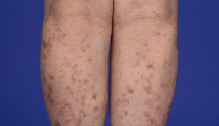
Lesions, or damaged areas on the skin, often take the form of ring-shaped red or brown patches that are sometimes itchy. These patches, which can take the form of flat spots, small bumps, or raised areas, have a thickened border. They usually occur in pairs. The lesions are more common in people in their thirties or forties and mainly appear on sun-exposed parts of the body. The legs, forearms, shoulders, and back are more prone to be affected. On rare occasions, they can appear on the face. However, the palms of the hands and the soles of the feet are usually unaffected. These lesions, which are associated with a condition called disseminated superficial actinic porokeratosis, typically worsen when exposed to sunlight, and the itchiness can also increase.
Testing for Disseminated Superficial Actinic Porokeratosis
Disseminated superficial actinic porokeratosis, a skin condition, can usually be identified by the look of the skin lesions. If there’s uncertainty about the diagnosis, a skin biopsy can be done. Using a tool called a dermoscope can also be helpful in examining this skin condition.
According to research by Nicola and colleagues, there are certain things that can be typically seen under a dermoscope when examining porokeratotic skin lesions:
- A white area around the whole lesion,
- A white area in the center of the lesion that looks like a scar,
- Small brown spots or dots,
- Blood vessels that are either little pinpoint vessels or irregular lines crossing through the lesion.
Treatment Options for Disseminated Superficial Actinic Porokeratosis
For conditions like actinic keratosis and disseminated superficial actinic porokeratosis, several treatments are available.
Topical diclofenac, a type of nonsteroidal anti-inflammatory drug (NSAID), can help manage this condition. It works by blocking a molecule called COX-2 and is considered safe for use. However, it yields different results in different patients.
Ingenol mebutate is another treatment option. It treats visible symptoms like skin thickening (hyperkeratosis), but can’t treat skin thinning (atrophy) or loss of skin color (hypopigmentation).
Vitamin D3 analogs, similar in function to the vitamin D our bodies produce, can help with this condition. Using it for 6 to 8 weeks can show good results. These analogs work by promoting the growth and development of skin cells (keratinocytes).
5-fluorouracil is a medication that works by inhibiting fast-growing cells. It can cause inflammatory reactions such as redness, ulcers, and dermatitis in some patients. Though its use can offer relief, the effects are usually temporary.
Imiquimod stimulates the body’s immune system to fight the condition. It can also cause an inflammatory response.
Photodynamic therapy is another choice for treatment. This involves applying a photosensitizer, which is a substance taken up by abnormal skin cells. When light is applied, the photosensitizer produces reactive oxygen species that destroy the abnormal cells. Two different photosensitizers, 5-aminolevulinic acid (ALA) and methyl aminolevulinate (MAL), can be used. Some studies suggest MAL might be more effective because it can penetrate the skin better.
Retinoids, derivatives of vitamin A, are used in conditions where there’s abnormal growth of skin cells. Topical use is preferred because intake of retinoids can cause more side effects and can be harmful during pregnancy. However, symptoms may return after treatment stops.
Cryotherapy (using cold to destroy tissue), surgical methods like excision and dermabrasion, and curettage (scooping out tissue with a curette) may be effective but are generally used for limited disease. These methods may leave scars, and the condition could recur.
Various types of lasers can treat this condition, each working in a different way to target and destroy abnormal skin cells. However, different lasers can leave different side effects, such as dark spots on the skin from the carbon dioxide laser.
Immunosuppressive agents, which reduce the body’s immune response, are not typically effective because disseminated superficial actinic porokeratosis is not an inflammatory disease. These medications can help with itching associated with the condition.
Finally, a combination of 2% simvastatin or lovastatin with 2% cholesterol cream can help decrease the number of skin lesions, scaling, and redness.
What else can Disseminated Superficial Actinic Porokeratosis be?
- Sunspots (Actinic keratoses)
- Ring-shaped lumps (Granuloma annulare)
- Reddish-purple skin rash (Lichen planus)
- Age spots (Seborrheic keratoses)
- Ringworm (Tinea corporis)
- Flat warts


