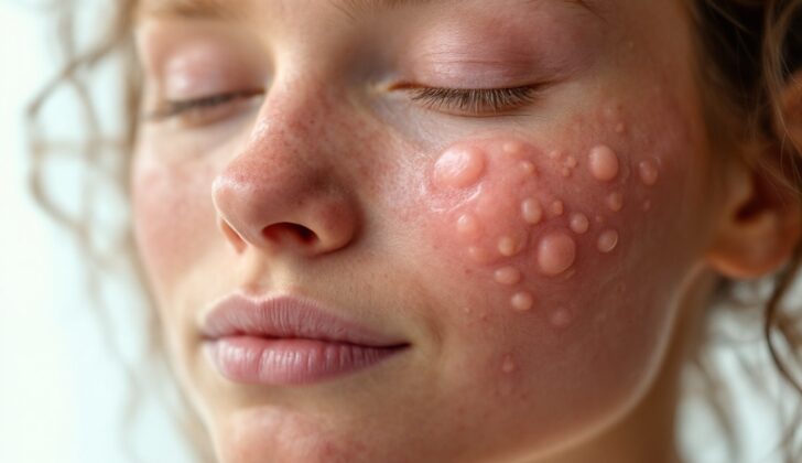
What is Epidermoid Cyst ( Sebaceous Cyst)?
An epidermoid cyst, often called a sebaceous cyst, is a harmless, enclosed bump under the skin filled with a protein called keratin. These cysts most often show up on the face, neck, and chest, but they can turn up anywhere on the body, including sensitive areas like the genitals, fingers, and the inside of the mouth. They tend to grow slowly and can linger for years.
The term ‘sebaceous cyst’ is frequently used, but it’s a bit misleading as it doesn’t involve the sebaceous gland, which is a small oil-producing gland in the skin. Instead, epidermoid cysts actually develop in the infundibulum, a part of the hair follicle. They’re also known as infundibular cysts, epidermal cysts, and epidermal inclusion cysts.
While these cysts are generally harmless, in very rare cases, they can develop into a cancerous growth.
What Causes Epidermoid Cyst ( Sebaceous Cyst)?
Most cases of epidermoid cysts, which are skin bumps that are typically non-cancerous, occur randomly. Sometimes, they are found in people with certain genetic conditions like Gardner syndrome and Gorlin syndrome, which are passed down through families. If a child gets these cysts in unusual places or in large numbers before puberty, it could suggest the presence of a syndrome.
Epidermoid cysts can also occur due to severe sun damage in older patients, a condition known as Favre-Racouchot syndrome. Certain medications like BRAF inhibitors, which are commonly used to treat certain types of cancer, can cause epidermoid cysts to develop on the face. Also, some recent findings show that medications like imiquimod and cyclosporine, which are used to treat a variety of conditions from skin cancer to psoriasis, can lead to the development of epidermal inclusion cysts, a type of cyst that’s similar to epidermoid cysts.
Risk Factors and Frequency for Epidermoid Cyst ( Sebaceous Cyst)
Epidermoid cysts are the most common kind of skin cysts, and they usually show up when people are in their 30s and 40s. It’s pretty rare to see these cysts before someone’s gone through puberty. More males have these cysts than females, with a ratio of 2 to 1. When babies are newborns, they often have small versions of these cysts, which are called milia. Around 1% of these cysts can turn into either squamous cell carcinoma (SCC) or basal cell carcinoma (BCC), which are types of skin cancer.
- Epidermoid cysts frequently appear in the 30s and 40s and are the most common type of skin cysts.
- Usually, these cysts are not found before puberty.
- They are more common in males than in females, with a ratio of 2:1.
- Newborn babies often develop small epidermal cysts known as milia.
- About 1% of these cysts can develop into squamous cell or basal cell skin cancers.
Signs and Symptoms of Epidermoid Cyst ( Sebaceous Cyst)
An epidermoid cyst is a type of small bump that can appear on various parts of the body. They can range anywhere from 0.5 cm to several centimeters in size and often have a firm, compressible feel. At the center of these cysts, there’s usually a dark opening, also known as a comedone opening or punctum. Often, these cysts don’t cause any discomfort or symptoms. However, if they rupture, they can become painful and resemble a boil, with redness and swelling. Additionally, a yellowish, cheese-like substance with a foul odor can sometimes be discharged from the ruptured cyst.
Interestingly, these ruptures can be caused by a sudden impact to the cyst, such as a person falling on their back or being slapped on the back. These cysts can occur virtually anywhere on the body, but the most common locations include the face, neck, chest, upper back, scrotum, and genitals. They can also be found on the buttocks, palms, and the soles of the feet, particularly if an injury has occurred in these areas. Distinct changes to the fingernails can sometimes be observed if the cyst appears on the end of a finger. Knowing the patient’s history, including any relevant injuries, medications, or genetic syndromes they may have, can provide important clues for confirming the diagnosis.
Testing for Epidermoid Cyst ( Sebaceous Cyst)
The assessment of epidermoid cysts, which are small, bump-like growths under the skin, largely depends on the person’s medical history and physical exams conducted by a doctor. There’s an ongoing discussion among health professionals about whether it’s required to analyze these cysts under a microscope after they’ve been removed.
Tests like bloodwork or imaging (like x-rays or ultrasounds) aren’t typically needed to check for these types of cysts.
Treatment Options for Epidermoid Cyst ( Sebaceous Cyst)
The best way to treat a cyst is through a surgical procedure that removes the entire cyst, including its wall. However, if the cyst is currently infected, it’s best to delay this complete removal. That’s because the inflammation from the infection can make it hard for surgeons to see the lines they need to cut along. If this is the case, doctors may decide to make an initial cut to drain the cyst, although this could mean the cyst might come back later on.
When removing a cyst, a local anesthetic, which numbs the area, combined with epinephrine (a medication that helps control bleeding), is often the best choice. The anesthetic is usually injected around the cyst and not directly into it. Doctors can then make a small, elliptical cut that includes the center of the cyst (known as the “central core” or “punctum”). It’s also crucial to align the cut with the natural lines of the skin to achieve the best cosmetic results. After the cyst is removed, the doctors will close the incision with multiple layers of stitches beneath the skin surface to achieve the best healing outcome.
Another method to remove the cyst could involve using a tool to take a small sample (a “punch biopsy”) and then gently push the cyst out through the small opening. And if the cyst is surrounded by inflamed tissue, a medicine known as triamcinolone may be used to reduce this inflammation. This might also mean delaying the surgical removal until the inflammation has reduced.
If the cyst has broken open on its own and its lining is destroyed, it usually will not come back. But it’s important to remove all fragments of the cyst’s lining during surgery to reduce the chance of it reappearing.
What else can Epidermoid Cyst ( Sebaceous Cyst) be?
When looking at the possible diagnoses for a condition known as an epidermoid cyst, doctors might consider several other conditions based on where in the body the cyst is located. These other possibilities include:
- Lipoma
- Dermoid cyst
- Pilar cyst which may also be known as an isthmus-catagen cyst, trichilemmal, or wen
- Furuncle
- Branchial cleft cyst
- Milia
- Pilonidal cyst
- Calcinosis cutis
- Pachyonychia Congenita
- Steatocystoma
- the skin-related symptoms of a condition known as Gardner syndrome
What to expect with Epidermoid Cyst ( Sebaceous Cyst)
Epidermal inclusion cysts are generally harmless. However, there’s a rare chance they could turn into a malignancy, which is a type of cancer. Two common types of malignancy that can develop from these cysts are Squamous cell carcinoma (SCC) and Basal cell carcinoma (BCC). These are types of skin cancers, with SCC being the more common of the two, occurring about 70% of the time.
Possible Complications When Diagnosed with Epidermoid Cyst ( Sebaceous Cyst)
If a cyst ruptures, it can lead to skin redness, swelling, and discomfort. If the cyst is surgically removed, complications may include bleeding, infection, and visible scars. Skin redness and pain can be managed with a medicine called ‘intralesional triamcinolone.’ Infections after surgery can be prevented by following proper sterile procedures. Although generally harmless, in rare cases, the occurrence of cancer in a cyst is possible.
Possible Complications:
- Skin redness, swelling, and discomfort after cyst rupture
- Bleeding, infection, and visible scars after surgical removal
- Potential for rare cases of cancer in a cyst
Recovery from Epidermoid Cyst ( Sebaceous Cyst)
After having surgery, it’s a good idea to steer clear of rigorous activities and sports that involve a lot of physical contact. The stitches from the surgery can usually be taken out after about a week to ten days. Doctors want patients to understand that the scar left from the surgery will take about two months to reach approximately 80% of the original skin’s strength. In case the scar needs to be improved or revised, this should be done between 6 months to a year after the surgery. This is because the remodeling phase of the wound healing, which is when your body continues to change and strengthen the scar, happens between 3 weeks to a year post-surgery.


