What is Granuloma Faciale?
Granuloma faciale (GF) is a rare skin condition that is not harmful but causes inflammation. It often appears as single or multiple patches on the skin that are well-defined, painless and can range in color from red-brown to a purple hue. These patches might also show accentuated hair follicles and tiny, widened blood vessels, known as telangiectasia. The term ‘granuloma faciale’ was first used by Wigley JE in 1945, who described the condition as ‘eosinophilic granuloma’.
Commonly, granuloma faciale is seen in white men who are in middle age (between 20 and 70 years old). However, there are instances of this condition appearing in children as well. These lesions usually occur on the face, with the forehead, nose, or cheeks being the most common locations. Although primarily a facial condition, granuloma faciale can affect other parts of the body like the scalp, trunk, inside the nose, or limbs.
What Causes Granuloma Faciale?
The exact cause of granuloma faciale, a skin condition, isn’t known. But, since it often appears on parts of the body that get a lot of sun, it might be linked to exposure to sunlight and the possible skin damage that can result from it. There are other factors that we think could contribute to this condition, like allergies, injuries, or even radiation therapy.
Risk Factors and Frequency for Granuloma Faciale
Granuloma faciale is a fairly uncommon condition predominantly found in adults. It affects both men and women, although it is slightly more frequent in men. It can occur in any race, but is most commonly seen in white individuals. Typically, the condition begins to show around the age of 52.
Signs and Symptoms of Granuloma Faciale
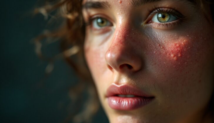
Granuloma faciale, a skin condition, usually shows up as a well-defined, painless bump or spots on the skin that are reddish-brown or purplish in color. These spots often have hair-like structures and small blood vessels on their surface. The most common places for granuloma faciale to appear are on the face and other areas exposed to the sun. A tool called a dermoscope can be helpful for doctors to examine these spots more closely. This tool shows certain features of the spots that include:
- Linear, tree-like blood vessels
- Enlarged openings of hair follicles
- Brown dots or globules, which are thought to be a sign of blood breakdown inside the skin
Testing for Granuloma Faciale
Granuloma faciale is a skin condition that gradually gets worse over time and frequently goes through periods of increased severity. It doesn’t have separate phases of being acute (short-term and severe) or chronic (long-lasting), making it difficult for doctors to assess how long it’s been present based on tissue sample studies. These studies often show features of both short-term and long-term inflammation.
Apart from slightly raised levels of eosinophils (a type of white blood cell) in the blood, tests usually come back normal. Granuloma faciale itself is not harmful, but it rarely goes away on its own; usually, treatment is required. However, the treatments we currently have often come with a high risk of the condition coming back.
Treatment Options for Granuloma Faciale
Granuloma faciale is a skin condition that can be tricky to treat, as results can vary from person to person. There are several potential methods of treatment that doctors may recommend.
First, there’s topical treatment, which means using creams or ointments applied directly to the skin. This is usually the first choice for treating granuloma faciale. Topical treatments include steroid creams (corticosteroids) or a medication called tacrolimus. A type of cream containing an antibiotic called dapsone is also sometimes used.
Another option is the use of intralesional steroids. This involves injecting steroids directly into the affected skin area. Some patients have reported positive results from this method.
There’s also the possibility of using systemic treatment, which means taking medication that treats your whole body. This could involve taking corticosteroids or dapsone in pill form.
Phototherapy, or light therapy, is another option. This involves exposing the skin to specific types of ultraviolet light and is sometimes done using a treatment process called psoralens plus ultraviolet A (PUVA).
There are other treatment methods also available, such as using lasers, cryotherapy (which means freezing the affected skin), or even having surgery to remove the affected area. The most suitable treatment for each patient will depend on their individual circumstances and the severity of their condition. The best course of action should be discussed with a healthcare provider.
What else can Granuloma Faciale be?
There are several skin conditions that can look similar to granuloma faciale. These may include:
- Sarcoidosis
- Discoid lupus erythematosus
- Rosacea
- Mycobacterial infections
- Cutaneous deep fungal infections
- Cutaneous lymphoma
- Basal cell carcinoma
Erythema elevatum diutinum (EED) can also be mistaken for granuloma faciale because it shows similar features under a microscope, like fibrosing vasculitis, which is a type of inflammation of the blood vessels. It’s thought that EED and GF might be caused by the same underlying issue. The major distinction is that EED generally appears as multiple lesions on the joints, whereas GF is usually a single lesion on the face.
Diagnosis can be difficult when EED shows up on the face or if GF appears somewhere other than the face. A high number of a type of white blood cell called eosinophils in the tissue can suggest a diagnosis of GF. Also, the presence of granulomatous nodules (clusters of immune cells) in the tissue can be seen in some cases of EED.
Lastly, EED is often linked with other underlying conditions like blood disorders, autoimmune diseases, HIV infections, other infectious diseases, and insect bites. However, GF is rarely associated with other systemic (whole-body) illnesses.


