What is Hypopigmented Macules?
Hypopigmented macules are common skin spots that have less pigment, or melanin, than your normal skin. They shouldn’t be mistaken for depigmented spots, which do not have any melanin at all because the cells that produce melanin, called melanocytes, have significantly declined. However, it can be hard to tell the difference between the two types of spots just by looking.
These two conditions must be distinguished because the treatment, as well as their impact on your health, can differ. Hypomelanosis, another term for the condition of hypopigmented spots, is mostly harmless, but can occasionally be linked with the function of internal organs, and very rarely with cancer. One of the main reasons people seek treatment is due to their appearance, which can create emotional distress and social discomfort, particularly in individuals with darker skin. Through accurate diagnosis and prompt treatment, some cases can regain their pigment. A thorough medical history, clinical signs coupled with a special lamp exam, and skin evaluation can all aid in diagnosing the condition correctly. Also, the study of a skin tissue sample under a microscope can provide additional clues about the nature of the disorder. This article will largely discuss the general diseases tied to hypopigmented skin spots that are smaller than 0.5 cm and patches larger than 0.5cm.
To understand these disorders of skin pigment, it’s pivotal to have a basic understanding of the skin’s structure and how skin pigmentation works. The skin is the body’s biggest organ and consists of three layers – the outer layer (epidermis), the middle layer (dermis) and the lower layer (subcutaneous or “hypodermis” layer). The epidermis has five sub-layers, the deepest of which is the basal layer. The most superficial layer, the stratum corneum, generally doesn’t contain any cell nuclei and is the layer usually visible.
Skin color is primarily dictated by two types of cells in the base layer of the epidermis: keratinocytes and melanocytes. These melanocytes originate from cells of the nerve tissue and their movement to the skin and hair is regulated by various internal factors and procedures. Melanocytes produce a brown pigment, melanin, which shields the skin from the harmful UV radiation in sunlight. Being exposed to UV light could cause changes in the DNA of keratinocytes, leading to the production more POMC protein. This protein turns into ACTH and alpha MSH which bind to the G-protein receptor (MC1R) on the melanocyte leading to the production of eumelanin, tyrosinase, and tyrosinase-related protein. The tyrosinase enzyme then transforms tyrosine into DOPA and melanin, which gives skin its color.
As we age, the keratinocyte cells from the base layer move up to the surface of the skin along with the melanin, which provides a brownish tint to the skin. There are two types of melanin: pheomelanin, which is predominant in fair-skinned people and eumelanin, more prevalent in dark-skinned people. Eumelanin offers better protection against UV radiation compared to pheomelanin.
What Causes Hypopigmented Macules?
Conditions where the skin becomes lighter, known as hypopigmented conditions, are mostly acquired or developed later in life. They can be rarely present from birth, which we call congenital conditions.
The congenital conditions include:
* Albinism – inherited from parents where both parents pass on the trait.
* Piebaldism – inherited from parents with one parent passing on the trait.
* Hypomelanosis ito – this occurs due to chromosome defects.
Conditions that are acquired or developed later in life, include:
* Normal aging – Idiopathic guttate melanosis (a condition characterized by small round spots on the skin that are lighter than the surrounding skin).
* Environmental factors like too much sun exposure and microtrauma – again, they lead to Idiopathic guttate melanosis.
* Nutritional deficiencies – these include Kwashiorkor (a severe lack of protein), vitamin B12 deficiency, and lack of copper and iron.
* Inflammatory causes – Pityriasis alba (light-colored patches on the skin that often relates to a skin condition called atopic dermatitis).
* Vascular causes – These include Bier’s spots, which are light spots that appear on the skin due to reduced blood flow.
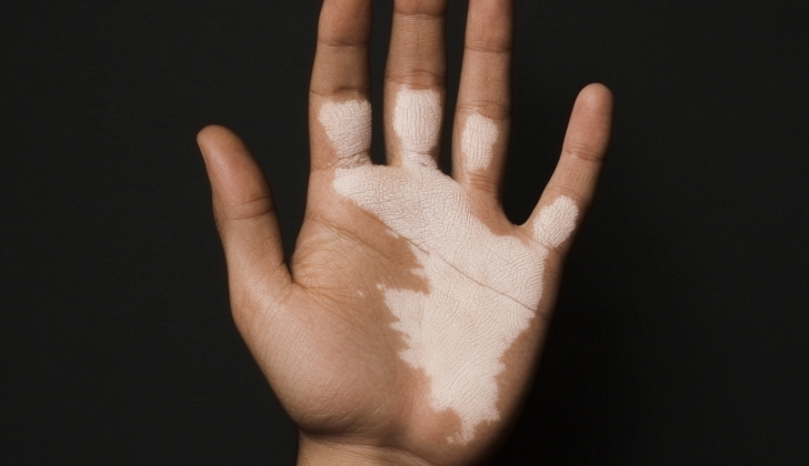
* Autoimmune causes – These include Vitiligo, a condition where some skin cells stop producing pigment leading to white patches on the skin, and hypopigmented sarcoidosis, where lighter patches appear on the skin due to underlying inflammation.
Several infections can also result in lighter skin, such as fungal infections by Malassezia species, bacterial infections like progressive macular hypomelanosis and leprosy, protozoal infections like post-kala-azar dermal leishmaniasis, and certain viral infections.
Chemical inflammation caused by products such as lead-based cosmetics and certain skin bleaching agents can also lead to lighter skin. Furthermore, inflammation due to any skin conditions, infections, or procedures (for example, cryotherapy or dermabrasion which are skin treatment procedures) can also lead to lighter skin. Lighter skin can also result due to burns or various other reasons. Certain types of cancers, like hypopigmented mycosis fungoides, a type of skin cancer, also result in lighter patches on the skin.
Lastly, certain inherited conditions like Ash leaf spots in tuberous sclerosis can also cause hypopigmented skin conditions.
Risk Factors and Frequency for Hypopigmented Macules
Hypopigmentation macules, or spots with less pigment, appear frequently in both children and adults, affecting at least 1 in 20 people. The occurrence of different skin conditions related to hypopigmentation may vary based on a person’s age, gender, race, geographical location, family history, and exposure to environmental factors.
- For instance, P. alba affects up to 90% of children under the age of 16.
- P.versicolor is more common in adolescents and young adults.
- IGH is prevalent in 80% to 87% of adults over 40 years old.
- Vitiligo, on the other hand, can affect anyone, regardless of age, from toddlers to the elderly.
- P.alba is slightly more common in males, but P.versicolor affects both genders equally.
- Hansen’s disease, also known as leprosy, is more common in developing countries, with immigrants making up more than 75% of cases in the United States.
- P.alba is typically seen in people who have a family history of atopic dermatitis, a type of skin condition that causes red, itchy rashes.
Signs and Symptoms of Hypopigmented Macules
Diagnosing different conditions that cause loss of skin color (hypopigmentation) can’t always be done just by looking at the changes in the skin. To find the cause and how serious the condition is (whether it was inherited or developed, and if it is harmless or harmful), doctors need a detailed patient history. This includes personal information, job-related information, and family medical history. Sometimes, patients might also experience symptoms that aren’t tied to their skin. A full physical exam, from head to toe, is essential to make the correct diagnosis. Informations gathered from a questionnaire and particular findings in a medical exam can be helpful for evaluating patients with light-colored spots on their skin.
- Did the skin changes appear at birth, or did they develop later in life?
- If they developed later, did this happen suddenly or gradually?
- Has there been any previous inflammation, or exposure to harmful chemicals?
- Are the lighter skin patches in one area or spread out?
- Where are the patches located? Are they on areas exposed to the sun, or in folds of the body like under the arm or below the breast?
- What do the patches look like? Factors to consider include:
- Size of the patches
- Are the edges defined or not?
- Are they scaly or smooth?
- What is their pattern?
- Do they affect one side or both?
- Are they stable or getting worse?
- Are they joining together or changing color?
- Do they itch or have scratches from itching?
- Has there been any previous inflammation, or exposure to harmful chemicals?
- Did the characteristics of the patches change according to weather variations?
- What does the skin around the patches look like?
Testing for Hypopigmented Macules
When trying to accurately diagnose a skin condition, a doctor will use an organized approach.
One method is the Wood’s lamp examination; this involves using a special lamp in a dark room to better see skin areas that have changed colour, especially those that have discoloured or lost pigmentation completely. The special lamp used, known as a Wood’s lamp, shines a type of ultraviolet light onto your skin. It is held 4-5 inches away from your skin. Healthy skin does not respond to this light, but skin affected by diseases such as vitiligo will appear bright white. This lamp can also identify certain skin infections by the colour they glow under the light.
Dermatoscopy is another technique used by doctors. This is basically a microscopic examination of the skin. This gives a detailed view of the skin abnormalities like any changes in pigmentation, the exact edges of the lesion, scaly or non-scaly appearance, any blood vessels close to skin surface and what the skin surrounding the discoloured patch looks like.
A KOH mount of skin scrapings is a quick test used to identify fungal infections like Tinea versicolor. The doctor will scrape some skin off and mix it with KOH, a strong solution, then examine it under a microscope. This helps to spot any sign of fungal infection.
In some cases, when the diagnosis isn’t clear or the doctor suspects an infectious disease or underlying medical condition like cancer, a skin biopsy may be necessary. This involves taking a small piece of skin tissue and examining it under a microscope using various stains. This can provide further detail about the condition and can be particularly helpful if the cause of the skin discoloration is unknown.
And finally, electron microscopy is another, less commonly used method, that can be used in certain settings. It provides an extremely detailed view of the skin lesion and helps differentiate the causes for loss of skin pigment. However, this technology is not commonly available and doctors usually have to rely on other clinical findings.
Treatment Options for Hypopigmented Macules
Hypopigmented macules, which are lighter patches of skin, are addressed based on what’s causing them in the first place. Often it’s possible to return the skin’s color back to normal by diagnosing the problem early, eliminating anything that might be causing it (like certain chemicals or infections), not exposing your skin to sunlight, using sunscreen, and getting appropriate treatment. However, if these skin patches are a result of inherited conditions linked to genetic defects, it might not be possible to restore their color. Treatments can speed up the process of returning color to the skin. There are several ways to do this, including medications, light therapy, and sometimes surgery.
For medications, there are topical (applied to the skin) and systemic (affecting the whole body) options.
Topical corticosteroids are often the first choice for many skin conditions causing loss of color. These medications can speed up the return of color to the skin. Sometimes, these are used in combination with light therapy to achieve better results. Systemic corticosteroids, which work throughout the body, are seldom used to manage discoloration, but may be used to stop the fast progression of skin lesions in certain diseases, like vitiligo.
Another kind of topical medication is called calcineurin inhibitors, which include tacrolimus and pimecrolimus. These are also first-line medications, meaning they’re among the first treatments doctors try. These drugs help protect the pigment-producing cells in the skin from harmful inflammation. Unlike topical corticosteroids, tacrolimus does not cause skin thinning and stretch marks, so it’s often preferred for treating facial skin discoloration.
Vitamin D appears to be linked with skin pigmentation. Vitamin D can increase the production of pigment by enhancing the functionality of special cells in skin called melanocytes. Antifungal medications are used when the skin lightening is due to a fungal infection called Tinea versicolor. Topical antifungals are usually applied for weeks or months and include medicines such as selenium sulfide shampoo and 1% or 2% ketoconazole ointment. Oral medications like fluconazole and itraconazole are used for a shorter period. Furthermore, oral isotretinoin can be used for conditions like PMH where a type of bacteria known as propionibacterium acne is the underlying cause. Lastly, moisturizers or emollients can soothe itchy and scaly skin patches.
Light therapy includes narrow-band ultraviolet B (NBUVB) and psoralen ultraviolet A (PUVA) treatments. NBUVB is generally considered better and is preferred over PUVA for treating vitiligo, as it has fewer side effects. PUVA, which uses a substance called psoralen, is not recommended for children or pregnant women due to its potential adverse effects.
In some cases, surgical procedures like skin grafting and split skin grafting can be performed to treat localized or inherited skin discoloration.
What else can Hypopigmented Macules be?
If you see light patches on your skin, they might be due to several conditions. Some possible causes include:
- P. alba, common in children and often linked with a history of skin irritation
- P. versicolor, usually seen in teens in hot, humid climates, and frequently appearing on seborrheic areas like the trunk, neck, and arms
- IGH, more prevalent in the elderly and often associated with prolonged sun exposure and repeated minor skin injuries
- Vitiligo, which causes loss of skin color in patches
- PMH, or post-inflammatory inflammation, which happens after the skin is injured or irritated
- Halo nevus, or a mole surrounded by a ring of lighter skin
Making a correct diagnosis among these conditions is important, as treatments and outcomes can differ. Information on your personal and family health history, physical examination, and the patterns of light patches on your skin can be helpful in identifying the cause. Certain characteristics may indicate the condition. For example, residual pigmentation around hair follicles is often seen in vitiligo but not in other conditions. Care should be taken to differentiate vitiligo from conditions like chemical leukoderma and skin lightening in cancer patients undergoing immunotherapy.
Hypopigmented mycosis fungoides (HMF), a rare form of skin cancer that often appears as light patches, may be overlooked, especially in children. This condition can be mistaken for other causes of skin lightening, such as eczema, vitiligo, and PMH. A high degree of suspicion and a skin biopsy are needed to confirm a diagnosis of HMF.
What to expect with Hypopigmented Macules
Most conditions related to hypopigmentation, or loss of skin color, are not harmful and usually have a positive outlook. Prompt identification and treatment often allows the skin to regain its color. However, inherited conditions, meaning those passed down through families, may not have as positive an outlook.
The outlook for conditions of hypomelanosis, which is a specific type of pigment loss, that are linked to underlying cancers varies. The timing of diagnosis is crucial, as these conditions are often misidentified as harmless ones such as Pityriasis Alba (a type of skin disorder), or Idiopathic Guttate Hypomelanosis (a condition that causes small white spots), which can lead to delayed treatment.
Possible Complications When Diagnosed with Hypopigmented Macules
People with lighter skin patches, or hypopigmented macules, are more at risk of skin cancer. This is due to less skin pigment, or melanin, in these areas, making them more exposed to the damaging effects of the sun’s UV radiation on skin cells. To lower this risk, it is recommended to minimize sun exposure and regularly apply sunscreen.
Diagnosing the actual cause behind the skin patches can also help in treating the overall disease more effectively instead of just focusing on the symptoms. For example, ash leaf spots on the skin can indicate the presence of a neurocutaneous disorder, such as tuberous sclerosis.
The psychological impact of having hypopigmented skin lesions can’t be ignored. It may lead to anxiety, stress, and depression in some individuals. Hence, psychological support and understanding may be needed alongside treatments for the skin condition.
Key Points
- Lighter skin patches can indicate higher risk of skin cancer
- Reducing sun exposure and using sunscreen can mitigate this risk
- Identifying the root cause of the skin patches can help in effective treatment
- Ash leaf spots on skin may signify a neurocutaneous disorder
- Psychological impact of having lighter skin patches can induce anxiety, stress, and depression
Preventing Hypopigmented Macules
Often, individuals seek medical help due to the emotional distress caused by the appearance of skin abnormalities. It is crucial for them to be made aware that the medications used to treat these issues can have side effects such as thinning skin, development of stretch marks, and the appearance of small, broken blood vessels known as telangiectasis. Additionally, a medication called psoralen, often used in a type of treatment called PUVA, could potentially cause liver damage. Young women should also be alerted to the potential negative effects these medications could have on pregnancy.
It’s important to know that even after successful initial treatment, skin abnormalities could reappear. Being educated about what causes these relapses can help reduce their occurrence.
Finally, patients are advised to keep an eye out for any additional symptoms beyond the skin lesions. These could hint at larger issues, and should be reported to their doctor immediately if noticed.


