What is Keratoacanthoma?
Keratoacanthoma (KA) is a type of non-serious, fast-growing skin lump that usually measures 1 to 2 cm in size. It often looks like a small dome with a hard, scaly plug in the middle. Over time, the way this lump has been categorized has changed. Before 1917, it was considered as skin cancer. However, in the 1920s, it was called a wart or an overgrown skin gland cyst. Between 1936 and the 1950s, it was referred to as molluscum sebaceum.
This condition typically begins with a quick growth phase, after which the lump stops growing and may even shrink on its own. These lumps can also vary in nature depending on the subtype which includes solitary keratoacanthoma, subungual keratoacanthoma, mucosal keratoacanthoma, giant keratoacanthoma, keratoacanthoma centrifugum marginatum, generalized eruptive keratoacanthoma of Grzybowski, and multiple keratoacanthomas Ferguson-Smith syndrome.
Despite being classified as harmless, keratoacanthoma shares some microscopic characteristics with a type of skin cancer known as squamous cell carcinoma (SCC). Because of these similarities, keratoacanthoma often needs to be treated.
What Causes Keratoacanthoma?
Various factors can cause keratoacanthomas. These factors could include exposure to ultraviolet (UV) rays, contact with harmful chemicals, a suppressed immune system, use of particular medications (BRAF inhibitors), inherited genes connected to p53 or H-Ras mutations, exposure to certain viruses like human papillomavirus (HPV), or recent injuries or surgeries on the affected area.
Keratoacanthoma is often linked to certain genetic conditions. These include Muir-Torre syndrome and Ferguson-Smith syndrome, which are passed down through families. Similarly, it could be connected to xeroderma pigmentosum, which makes the skin highly sensitive to sunlight, incontinentia pigmenti, where the skin becomes discolored, and Witten-Zak, all of which can be genetically inherited.
A particularly unusual form of keratoacanthoma is known as ‘generalized eruptive keratoacanthoma of Gryzbowski.’ This condition involves widespread, itchy growths appearing all over the body.
Risk Factors and Frequency for Keratoacanthoma
Keratoacanthomas are skin conditions that can affect anyone, but are rare in people under 20. They are most commonly seen in people aged 50 to 69, and are twice as common in men than in women. They mainly affect people with fair skin, especially those with Fitzpatrick I-III skin type. Moreover, they are more common and tend to be more invasive in people with weakened immune systems. It’s also worth noting that keratoacanthomas share many characteristics with a type of skin cancer known as squamous cell carcinoma.
- Keratoacanthomas can affect all ages but are rarely seen in people under 20.
- The most common age range for this condition is between 50 and 69.
- Men are twice as likely as women to have keratoacanthomas.
- Fair-skinned individuals, particularly those with Fitzpatrick I-III skin type, are more likely to develop keratoacanthomas.
- Those with weaker immune systems experience these conditions more frequently and they tend to be more invasive.
- Keratoacanthomas share many features with squamous cell carcinoma, a type of skin cancer.

Signs and Symptoms of Keratoacanthoma
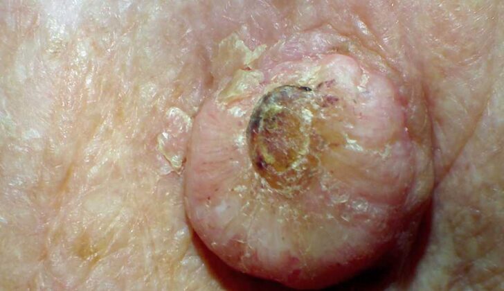
Keratoacanthoma is a skin lesion often found on parts of the body that are exposed to the sun, like the face, head, neck, and top of the arms and legs. Finding these lesions on the trunk of the body is less common. They start as small, pink or skin-colored bumps that grow rapidly into a dome-shaped lump with a unique central core that resembles a crater. These lesions typically measure between 1 to 2 cm in size.
Note that keratoacanthoma can also occur on areas that aren’t often exposed to the sun, such as the inner surface of the mouth, around the nails, buttocks, and anus.
During a physical examination, doctors usually check the surrounding lymph nodes due to the risk of the lesion spreading. Using a dermoscope to examine the lesion can help, but it doesn’t reliably differentiate between keratoacanthoma and another skin cancer called squamous cell carcinoma. However, it can help distinguish these from other non-pigmented skin bumps.
Particular signs to look out for under a dermoscope include blood spots, white circles, and the presence of keratin. Of these signs, white circles are the most specific clue in identifying this type of lesion.
Testing for Keratoacanthoma
If there are abnormal growths (also known as lesions), doctors will first ask you about your medical history and then do a physical examination. A biopsy, which is taking a small tissue sample from the body to examine in a lab, may be performed. This is important for a close investigation of the tissue.
The best test is something known as an excisional biopsy. This means the entire lesion (or abnormal growth) is removed and examined. This is done instead of a shave biopsy, where only part of the growth is removed, because it’s sometimes not enough to tell the difference between a type of growth called a keratoacanthoma and skin cancer (squamous cell carcinoma).
For those with keratoacanthomas under the fingernails or toenails (known as subungual keratoacanthomas), x-ray images (radiographic imaging) of the affected finger or toe are necessary. This is to check for osteolysis, which is the breakdown of bone caused by the growth.
Treatment Options for Keratoacanthoma
Keratoacanthoma is a skin condition that’s generally harmless. However, because it’s related to a type of skin cancer called squamous cell carcinoma, treatment is usually recommended. The first choice for treatment is a surgical procedure that removes the lesion or growth, making a cut about 4 millimeters from its edge.
If the growth is small, less than 2 centimeters, and on your arms or legs, a treatment called electrodessication and curettage might be used. This involves using electricity to dry out the skin growth, then scraping it away.
For larger growths over 2 centimeters that are located in areas of high cosmetic concern, a procedure called Mohs micrographic surgery may be considered. This procedure helps spare healthy skin while removing the growth.
Mohs surgery is also the primary treatment for keratoacanthomas that have developed in the nerve cells in the skin, a condition known as perineural invasion.
There are also some non-surgical treatments that have had limited use. These include applying creams containing imiquimod or fluorouracil (5-FU) directly to the skin. Other therapies have involved injecting the growth with drugs such as methotrexate, bleomycin, or 5-FU, or taking a drug called isotretinoin by mouth.
Out of the injected therapies, the most research has been done on 5-FU and methotrexate. They’re typically administered weekly for a period of 3 to 8 weeks. The data suggests these treatments have been effective at causing the growth to shrink in 83% to 100% of patients who received them.
What else can Keratoacanthoma be?
When checking for a skin issue that might be keratoacanthoma – a kind of skin tumor, doctors have several other conditions they need to rule out. These conditions include:
- Squamous cell carcinoma (a type of skin cancer)
- Amelanotic melanoma (another skin cancer type)
- Molluscum contagiosum (a skin infection caused by a virus)
- Prurigo nodularis (a skin disease causing itchy bumps)
- Metastatic lesions (areas where cancer has spread)
- Merkel cell carcinoma (a rare and aggressive skin cancer)
- Nodular basal cell carcinoma (a common form of skin cancer)
- Ulcerative basal cell carcinoma (an aggressive variant of basal cell carcinoma)
- Nodular Kaposi sarcoma (a skin cancer associated with AIDS)
- Hypertrophic lichen planus (a skin condition that results in bumps on the skin)
- Deep fungal infection (infections under the skin caused by fungi)
- Atypical mycobacterial infection (infections with organisms related to the ones that cause tuberculosis)
- Foreign body reaction (reaction to an alien substance in the body)
- Verruca vulgaris (commonly known as a wart)
This list shows that skin conditions can have similar symptoms but extremely different causes and treatments. It’s crucial for a proper diagnosis to be made so the right treatment can be provided.
What to expect with Keratoacanthoma
Keratoacanthoma, a type of skin condition, generally has a very good outcome after surgical removal. People who have keratoacanthoma usually have a history of exposure to the sun and it’s important for them to have regular check-ups to watch for any new skin cancers. While it’s rare for this condition to spread to other parts of the body (referred to as metastases), there have been some cases reported in medical literature where keratoacanthoma has spread along the nerves, a condition known as perineural spread.


