What is Langerhans Cell Histiocytosis?
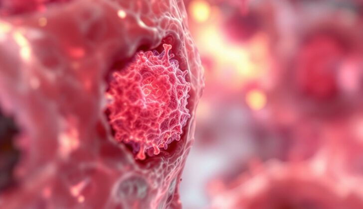
Langerhans cell histiocytosis (LCH) is the most common disorder that happens when specific cells, known as langerin-positive cells, come together and form lesions like small lumps in various parts of the body. These cells, essentially, cause inflammation in different tissues throughout the body. The areas affected can include places like the skin, liver, gastrointestinal tract (or the digestive system), and parts of the central nervous system (which includes the brain and spinal cord), such as the skull and pituitary gland (a small pea-sized organ in the brain).
Accurately diagnosing LCH is a team effort. It starts with a complete review by a pathologist, who is a doctor that studies diseases. They will examine the shape, chemical makeup, and molecular data of a tissue sample from the patient suspected of having LCH. Once LCH is confirmed, healthcare providers need to be aware that the treatment process can be different for children and adults. Particularly in adults, the kind of LCH (whether it affects the lungs or other parts of the body) plays a significant role in deciding the treatment plan.
Following a proper referral to a hematologist (a doctor who specializes in blood disorders) is crucial in order to manage LCH successfully. Regular check-ups and long-term follow-up with this specialist is a must.
What Causes Langerhans Cell Histiocytosis?
Langerhans cell histiocytosis (LCH) is a disease that scientists are still trying to understand fully. One popular theory is that it starts in a person’s bone marrow with the abnormal growth of specific cells called CD1a and CD207. These cells are early versions of blood cells, which turn into a type of white blood cell called monocytes. These monocytes then start to act like Langerhans cells, a type of cell involved in immune response.
Going deeper into this process, experts believe there might be a relationship between the unusual growth of these early blood cells (particularly the ones with changes in a gene called TET2) and changes in other genes (like BRAF) that can occur when monocytes develop. This essentially results in too many monocytes, which, when they enter the bloodstream, lead to Langerhans cell histiocytosis.
Risk Factors and Frequency for Langerhans Cell Histiocytosis
LCH, or Langerhans Cell Histiocytosis, is a rare disease. It mostly affects children under the age of 15, with the average age of diagnosis being 3 years old. It’s even rarer in adults and doesn’t occur often.
- LCH is rare, happening in up to 8.9 per million children under age 15.
- The average age a child is diagnosed with LCH is 3.
- This disease is even less common in adults, occurring in about 0.07 per million each year.
Signs and Symptoms of Langerhans Cell Histiocytosis
Langerhans cell histiocytosis (LCH) is a complex disease that can affect many parts of the body, including the skin, bones, lymph nodes, and internal organs like the lung, liver, and spleen. Depending on the area affected, people may experience different symptoms, and it can also vary greatly depending on a person’s age.
- In children, a rash is often the first sign. This can sometimes be mistaken for other skin conditions like seborrheic dermatitis. Yet, the LCH rash is unique – it could present as scaly patches, lumps, or plaques and may also include petechiae (small red or purple spots), bloody crusting, or hard lumps.
- Adults, especially those over 40 years old, usually display symptoms relating to multiple organ systems. Pulmonary LCH, which primarily impacts the lungs, is almost exclusively found in smokers and can cause symptoms like shortness of breath and coughing.
- When it comes to the liver, spleen, and hematopoietic system, clear definitions and criteria have been set for diagnosis. For example, liver or spleen involvement can be determined when these organs are enlarged beyond a certain size. If at least two types of blood cells are affected, it indicates hematopoietic involvement.
Determining the extent of the disease at diagnosis is critical in planning treatment. Healthcare providers must identify if a single system or multiple systems are affected.
The disease can also affect the central nervous system, causing a variety of symptoms based on the specific part of the brain involved. MRI scans are the go-to method for capturing images of the affected region, making neuroradiologists key in diagnosing this condition.
Testing for Langerhans Cell Histiocytosis
In 2022, a group of international experts on adult Langerhans Cell Histiocytosis (LCH), a rare type of cancer that can affect many parts of the body, presented some recommended steps for evaluating adult patients who may be suspected of having LCH or who have already been diagnosed with it.
To definitively confirm a diagnosis of LCH, it is most crucial to do a tissue biopsy (a procedure where a small piece of affected tissue is removed and analyzed). This is because the small changes under the microscope characteristic of LCH, such as cells staining positive for specific markers like S-100, CD207, and CD1a, can provide a decisive diagnosis.
It’s also important that doctors determine the genetic characteristics of the disease in every patient, specifically looking for mutations in the BRAF/MAPK/ERK genes. This is usually accomplished via specialist techniques like whole exome sequencing or integrated genomic-transcriptomic sequencing. These methods help in identifying other subtle genetic alterations that might be present.
While an electron microscope can be used to identify specific tiny structures within cells (known as Birbeck granules), which are a hallmark of LCH, this is usually not necessary due to advancements in staining techniques. It’s also recommended that tissue biopsy be performed in Pulmonary Langerhans Cell Histiocytosis (PLCH), a specific type of LCH affecting the lungs.
Once LCH is confirmed, imaging tools like a CT (Computed Tomography) scan or MRI (Magnetic Resonance Imaging) are suggested to visualize which organs are affected by the disease. For a more complete view of the disease, a certain type of scan, called a whole-body FDG-PET scan (which can show where cancer may be in the body), can be helpful.
Especially in cases where LCH overlaps with Erdheim Chester Disease (another rare type of disease), this scan can prove to be extremely useful. Should there be any suspicion of the disease affecting the hypothalamus or pituitary (parts of the brain), an MRI of the brain is also recommended.
Lastly, further specialized tests involving invasive or endoscopic procedures (procedures using a thin tube with a camera to view inside the body) are decided based on how the patient is doing and what symptoms they are showing.
Treatment Options for Langerhans Cell Histiocytosis
The management of Langerhans cell histiocytosis (LCH), a type of cancer that can affect different parts of the body, differs based on the extent and specific location of the disease. Here’s what you need to know:
1. Single-system unifocal LCH: This means that a single lesion (a damaged or altered area) is affecting one organ in the body. For this type of LCH, surgery is the most common form of treatment. The aim of the surgery is to entirely remove the lesion. There’s no need for additional drug treatments in concurrent use with the surgery. However, if the disease is affecting the skin, targeted radiation or injections of corticosteroids, such as triamcinolone, can be used.
2. Single-system pulmonary LCH: This refers to cases where the lungs are the only organ affected. Here, the most important thing is to stop smoking, including the use of devices like electronic cigarettes and marijuana, because there’s a known link between tobacco use and LCH. Quitting smoking can indeed induce complete remission. Medication such as cladribine may also be an option. Supportive therapies that focus on helping lung function—like beta-2 agonists and inhaled corticosteroids—are key.
3. Single-system multifocal LCH and Multisystemic LCH: This includes cases where more than one lesion is present but confined to a single organ or cases where more than two organs are affected. Treatment options are broader here and many are borrowed from techniques used to treat children with LCH. Among these options are drug regimens involving vinblastine and methotrexate, as well as cladribine used alone. When choosing a treatment plan, a provider will take into account the patient’s overall health and expected ability to tolerate the treatment.
Remember that each patient is different, and treatment plans may vary based on individual and specific circumstances. All recommendations mentioned above are adapted from international expert consensus guidelines.
What else can Langerhans Cell Histiocytosis be?
When diagnosing Langerhans cell histiocytosis (LCH), doctors need to consider other conditions that can have similar symptoms. These conditions include:
- Acrodermatitis Enteriropathica
- Acropustulosis of Infants
- Congenital Candidiasis
- Eosinophilic Pustular Folliculitis
- Incontinentia Pigmenti
- Leukaemia
- Lymphoma
- Mastocytosis
- Myeloma
- Neonatal Pustular Melanosis
Surgical Treatment of Langerhans Cell Histiocytosis
In the event that someone is diagnosed with a limited and singular disease, it’s crucial to involve a cancer surgeon as early as possible. The focus of the treatment would be to remove as much of the visible disease as possible during surgery, aiming for a near-complete removal if it can be achieved safely.
What to expect with Langerhans Cell Histiocytosis
The outlook of LCH, or Langerhans Cell Histiocytosis, can differ greatly because the disease can vary in how widespread it is and in its nature or phenotype.
Regrettably, nearly 50% of people with LCH are susceptible to a number of complications such as:
– Issues with muscles and bones
– Scarring of the skin
– Diabetes insipidus, a condition causing excessive thirst and urination
– Hearing problems
– Mental health issues like depression and anxiety, and cognitive issues
– Pulmonary or lung impairment
– Secondary cancers like lymphoblastic leukemia, which impacts the white blood cells, and solid organ tumors
– Slow growth rates in children
– Liver scarring, also known as cirrhosis
The long-term outlook for both kids and adults heavily depends on how far the disease has spread. According to one retrospective study (a study looking at past data), single-system LCH, meaning the disease is confined to one area or system in the body, often has a very good outlook – nearly 100% survival, with less than a 20% chance the disease will come back within 5 years.
However, if LCH involves multiple systems and organs at risk – like the spleen, bone marrow, or liver – the 5-year overall survival rate is equal to or less than 77%. Although this information comes from looking at past data, it does provide an insight into the real-world impact of LCH on patients.


