What is Milia?
Milia are non-harmful, short-lived skin cysts filled with keratin, a type of protein. They appear as small, hard, white bumps and can be found in varying numbers, mostly on the face. However, they can also show up on other parts of the body like the upper chest, arms and legs, and genital area.
Milia can be divided into two categories: primary and secondary. Most primary milia are congenital, which means they show up naturally and are present at birth. You can typically find them on the baby’s nose, scalp, eyelids, cheeks, gum line (known as Bohn nodules), and palate (called Epstein pearls). But in some cases, primary milia can also appear due to certain rare genetic skin disorders in both children and adults.
On the other hand, secondary milia develop due to an underlying skin condition, certain medications, or skin injuries. Regardless of the category, it’s important to know that milia are harmless and typically resolve on their own over time.
What Causes Milia?
Primary milia, a type of small bump or cyst, are thought to originate from the part of the hair follicle that is connected to oil glands in the skin. On the other hand, secondary milia, another type of small bump, are thought to come more often from sweat glands rather than from hair follicles, oil ducts, or the top layer of skin.
Primary milia have been connected to certain inherited genetic skin conditions. These include oro-facial digital syndrome type 1, a type of hair growth disorder called congenital hereditary trichodysplasia (also known as Marie-Unna hypotrichosis), Basex-Dupre-Christol syndrome, and some other skin disorders that cause abnormalities in the structures of the skin. A very uncommon form of primary milia that appears on a red patch of skin is known as milia en plaque. The cause of milia en plaque is still not well known.
Secondary milia can appear after skin injuries, such as from skin resurfacing procedures like dermabrasion, or from radiation therapy. They can also develop due to long-term use of topical steroids that thin out the skin, and from the use of nonsteroidal anti-inflammatory drugs. Also, they can occur after inflammatory skin conditions clear up, specifically in cases of blistering disorders.
Risk Factors and Frequency for Milia
Primary congenital milia, small, raised, white skin bumps, are incredibly common and can even be seen as typical. They occur in almost half of all healthy, full-term newborn babies. However, these bumps may take a bit more time to appear in premature babies.
There are two types, primary and secondary milia, and both can occur in people of any race or gender. The only exception is a type of milia called milia en plaque, which is more commonly found in females.
Milia can occur at any age but are most often seen in newborns that have the congenital form.
- Primary congenital milia is very common, seen in about 40% to 50% of full-term newborn babies.
- They might appear later in premature babies.
- Both primary and secondary milia can occur in anyone, regardless of their race or sex.
- The milia en plaque type is more common in females.
- While anyone can get milia, they are most common in newborns with the congenital form.
Signs and Symptoms of Milia
Milia is a skin condition that often appears at birth, but can also show up weeks later in premature babies. These small, usually painless, bumps will usually disappear on their own in the first few months of life. However, there are different types of milia, and they may show up differently.
For example, the acquired form of milia typically affects older children and includes a history of trauma or blistering skin diseases. This type of milia might not go away without treatment.
Milia is generally categorized into primary and secondary types as follows:
- Primary Milia:
- Congenital
- Benign primary milia of children and adults
- Milia en plaque
- Nodular grouped milia
- Multiple eruptive milia
- Nevus depigmentosus with milia
- Genodermatosis-associated
- Secondary milia:
- Disease-associated
- Medication-associated
- Trauma-associated
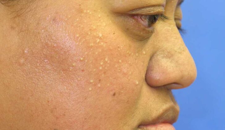
When it comes to identifying milia, they typically appear as small bumps less than 3 mm in size, varying from yellowish to white in color, and are usually found on the face, especially the nose. In older children and adults, milia often prefer the eyelids, cheeks, forehead, and genital area.
Some types of milia can present slightly differently. For instance, milia en plaque shows up as a reddened patch covered with multiple milia, which can be several centimeters in size. Multiple eruptive milia, another type, appear suddenly over weeks to months and can be widespread. It might be passed down in families, or be connected to certain genetic skin disorders. Milia has been known to occur alongside conditions like Brooke-Spiegler syndrome, pachyonychia congenita type 2, and basal cell nevus syndrome.
Milia can also associate with skin diseases like porphyria cutanea tarda or epidermolysis bullosa or occur due to using chronic topical steroids and nonsteroidal anti-inflammatory drugs. In other cases, they can appear after skin injuries like burns, skin grafting, dermabrasion, or radiotherapy.
Testing for Milia
Milia, also known as “milk spots,” is typically identified based on what a doctor sees during a physical examination. Sometimes, doctors may cut open one of these tiny bumps and examine the whitish, hard material inside to confirm their diagnosis. If the milia spots are widespread and stick around for a long time, the doctor might look into other potential causes of the spots, such as a genetic skin condition, especially if there are additional symptoms.
Treatment Options for Milia
Congenital milia, which are tiny, white, hard bumps that appear on the skin at birth, don’t usually need treatment. They tend to disappear on their own over time. However, other types of primary milia that develop later in life, and secondary milia, which occur after a skin condition or injury, may not go away without intervention.
If they don’t disappear naturally, they can be treated with minor surgical procedures. In these procedures, a small cut may be made with a scalpel (a very sharp surgical knife), and a tool called a comedone extractor or a curette could be used to apply sideways pressure to remove the milia.
Other treatment options for multiple milia involve using topical retinoids, which are creams or gels derived from vitamin A that helps promote skin turnover. Alternatively, a process called electrodesiccation or electrocautery could be used. In these treatments, an electric current is applied to the milia to dry them out or burn them off.
What else can Milia be?
When dealing with skin conditions, doctors often have to rule out other similar conditions to get a precise diagnosis. For example, if you have white to yellow grouped bumps around your upper lip and nose, you could be dealing with a condition called sebaceous hyperplasia. This condition is much like congenital milia (skin bumps you’re born with), but it’s not very common in babies born earlier than expected.
Other conditions that may have similar appearance include:
- Comedonal acne (a type of acne with blackheads and whiteheads)
- Flat warts (small, smooth, flat-topped skin growths)
- Milia-like idiopathic calcinosis cutis (a rare skin condition with small, hard, white bumps).
What to expect with Milia
Congenital milia, which are small, white bumps that occur on the skin at birth, usually clear up on their own within a few weeks, without leaving any scars. Sometimes, they can last for several months but usually disappear during a baby’s first month of life. Acquired milia, which form after birth, may persist without treatment.
Possible Complications When Diagnosed with Milia
There haven’t been any proven complications that affect the entire body. Milia, which are harmless and don’t cause any discomfort, are commonplace.
Key Facts:
- No systemic (whole-body) complications
- Milia are harmless and don’t cause discomfort
Preventing Milia
Doctors should talk to the parents or those looking after a child about the harmless nature of milia. Milia are tiny, white bumps that usually appear on a newborn’s face. The caregivers should understand that these bumps often vanish on their own, without leaving a scar.


