What is Necrobiosis Lipoidica?
Necrobiosis lipoidica is a rare, long-term, and unexplained skin disease which involves the breakdown of collagen, a protein important for skin structure. This causes skin sores, usually on the front side of the lower leg. The disease is often found in people who have diabetes, particularly type 1 diabetes, and can result in skin ulcers or open sores. The skin changes seen include thickening of blood vessel walls, breakdown of collagen, formation of small nodules of cells, and build-up of fat. The biggest problem with this condition is the development of ulcers, usually after an injury. Infections are rare. However, if necrobiosis lipoidica lasts for a long time, it can rarely become a type of skin cancer called squamous cell carcinoma.
Necrobiosis lipoidica was first acknowledged as “dermatitis atrophicans lipoidica diabetica” by a doctor named Oppenheim in 1929. But in 1932, another doctor named Urbach gave it the name ‘necrobiosis lipoidica diabeticorum’. The first case in a person without diabetes was reported by a doctor named Goldsmith in 1935. More cases in non-diabetic patients were described later by different scientists. In 1960, further cases in patients without diabetes were published, leading to a suggestion to remove the word ‘diabetic’ from the name. So, now, the broader term ‘necrobiosis lipoidica’ is used for all patients with these skin changes, whether or not they have diabetes.
Necrobiosis lipoidica is very commonly associated with diabetes, especially needing high amounts of insulin. But it’s also linked with metabolic syndrome (a group of conditions that increase your risk of heart disease, stroke, and type 2 diabetes), obesity, high levels of fat in the blood, high blood pressure, celiac disease (an autoimmune disorder where the ingestion of gluten leads to damage in the small intestine), and autoimmune thyroid disease. This suggests that there are other possible causes for the skin disease. Necrobiosis lipoidica is a disease with many potential factors contributing, and researchers are continually discovering new ways that might help treat it.
What Causes Necrobiosis Lipoidica?
While a large number of people with a skin condition called necrobiosis lipoidica also have diabetes, only a very small percentage of people with diabetes end up developing necrobiosis lipoidica. This suggests other factors also play a role in causing this skin condition. It’s believed that changes in blood flow, brought on by the depositing of immune substances or small blood vessel changes, can cause skin cell damage.
People with necrobiosis lipoidica may also have other health conditions. One study found that people with necrobiosis lipoidica were three times more likely to have thyroid disease, particularly autoimmune thyroid disease. Similarly, about a quarter of people with necrobiosis lipoidica also had thyroid disease, regardless of whether they had diabetes or not. High blood pressure was also common among people with necrobiosis lipoidica. Other often-seen health conditions included high cholesterol, coronary artery disease, obesity, metabolic syndrome, celiac disease, and other autoimmune diseases like sarcoidosis and multiple sclerosis.
There are many theories as to what causes necrobiosis lipoidica. One major theory involves diabetic microangiopathy, the damage to small blood vessels caused by diabetes, as a main cause of necrobiosis lipoidica. This is supported by the idea that the blood vessel damage seen in the eyes and kidneys of diabetics is similar to the changes seen in necrobiosis lipoidica. Additionally, a type of protein found in the blood vessel walls of people with this type of vascular damage is also seen in those with necrobiosis lipoidica.
Another theory suggests the depositing of certain immune substances, C3 and fibrinogen, in blood vessel walls could cause necrobiosis lipoidica. Some propose that a reaction initiated by an antibody called immunoglobulin M (IgM) could start the blood vessel changes that lead to necrobiosis lipoidica. These deposits are often located where the skin and under layer meet.
Abnormal collagen, a protein that provides structure to skin and other parts of the body, might be linked to necrobiosis lipoidica. Abnormal collagen fibers have been known to cause damage to various organs and speed up aging in people with diabetes. Higher levels of an enzyme that causes collagen to link together could possibly lead to the thickening of the skin’s lowest layer seen in necrobiosis lipoidica.
It’s also possible that defective white blood cells, specifically neutrophils, could cause an increased activity in a type of white blood cell called a macrophage. This could then lead to the formation of small inflamed nodules in the skin.
Lastly, a substance called tumor necrosis factor-alpha (TNF-α), which is crucial in certain diseases that involve these inflamed nodules, might play a role in causing necrobiosis lipoidica. Higher levels of TNF-α have been found in the blood and skin of patients with these conditions.
Risk Factors and Frequency for Necrobiosis Lipoidica
Necrobiosis lipoidica is a condition that often occurs in patients with diabetes mellitus, but this is not always the case. Among people with diabetes, only 0.3% to 1.2% will develop this condition. There are cases where the development of necrobiosis lipoidica may precede, coincide with, or follow the diagnosis of diabetes. However, there is no clear link between the control of blood sugar levels and the risk of this disease.
Necrobiosis lipoidica can also appear in healthy individuals, or those with other conditions like thyroid disorders and inflammatory diseases. Some of these diseases include Crohn’s disease, ulcerative colitis, rheumatoid arthritis, and sarcoidosis. We see this disease more frequently in females, especially in female diabetics, at a ratio of around 3 to 1.
- Necrobiosis lipoidica generally starts during the third decade of life (20s) in patients with type 1 diabetes, and in the fourth decade of life (30s) in people with type 2 diabetes or without diabetes.
- About one-third of patients will experience ulceration (sores). This is most common in men and patients with diabetes.

Signs and Symptoms of Necrobiosis Lipoidica
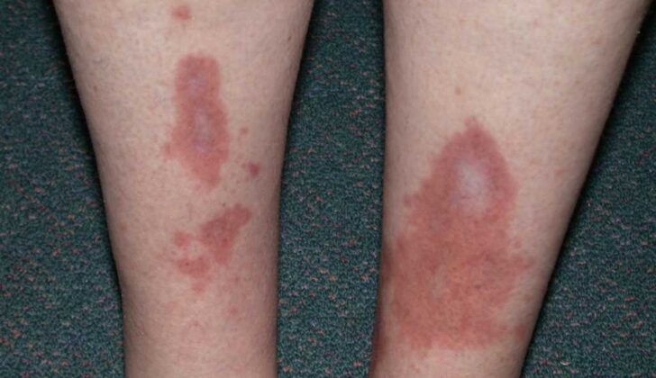
Necrobiosis lipoidica is a skin condition usually present on the front of the lower legs. It shows up as yellow-brown, thin, and patchy skin spots with a purple outline. In 85% of cases, these spots only appear on the lower legs, but they can show up in less common areas like the upper arms, face, scalp, chest, or penis.
The patches usually start as small, firm, red-brown bumps about 1 to 3 millimeters in diameter. These bumps gradually get bigger and merge into larger patches. The patches have hard edges and shiny centers. At first, the patches are red to brown but soon turn more yellow and thin out. They are often found as multiple patches on both sides of the body and can develop into ulcers, especially after minor scratches, in about one-third of cases. Interestingly, men tend to have worse symptoms compared to women, with ulcers reported in 58% of men compared to 15% of women. A study found that 17% of patients with this condition saw their ulcers heal on their own, but in most cases, the ulcers remain if untreated.
While the patches from this condition can cause the skin to be less sensitive to touch and painful, itchy, or produce a tingling sensation, some patients also report reduced sweating over these patches. Hair loss can also occur within the patches. On rare occasions, these patches can produce squamous cell carcinoma, a type of skin cancer. In addition, any trauma or surgery to the skin can lead to more patches.
Testing for Necrobiosis Lipoidica
If your doctor suspects you have a condition called necrobiosis lipoidica, they will order some blood tests. These tests can identify other diseases that often go hand-in-hand with necrobiosis lipoidica. One of these conditions is diabetes, so your doctor will test your fasting blood glucose or glycosylated hemoglobin. These measures show how high your blood sugar gets and how well it’s controlled. If these tests don’t confirm that you have diabetes, your doctor will repeat them every year since necrobiosis lipoidica can sometimes be the first sign of diabetes.
Because necrobiosis lipoidica is often associated with thyroid disease, your doctor may check your thyroid-stimulating hormone, free thyroxine levels, and thyroperoxidase antibodies. These tests can determine if your thyroid is working correctly.
If you have digestive symptoms or signs of celiac disease, your doctor may test for tissue transglutaminase, endomysial, and reticulin antibodies. If the doctor notices other symptoms of autoimmune disease, such as additional skin or joint symptoms, they might ask for evaluations for rheumatologic disease.
While your doctor can often diagnose necrobiosis lipoidica from a physical examination, they should also take a small sample of your skin (a biopsy). This helps to differentiate necrobiosis lipoidica from conditions with a similar appearance, like granuloma annulare and necrobiotic xanthogranuloma.
If your doctor is concerned that you may also have a vascular disease (conditions that affect your blood vessels), they might recommend further studies. This could include a vascular ultrasound, which uses sound waves to look at blood flow in your blood vessels, a computed tomography angiography, which is a type of X-ray that looks at your blood vessels, or an angiogram, which is a test that uses dye and special x-rays to show the insides of your arteries.
Treatment Options for Necrobiosis Lipoidica
As of now, there’s no definitive treatment for necrobiosis lipoidica, a skin condition. In some cases where the condition is not causing any pain or symptoms, doctors may prefer to simply monitor the condition over time. For people with swollen legs due to venous disease or lymphedema, which is a blockage in the lymph vessels, compression therapy can help manage the swelling and promote healing. Good wound care is essential when there are ulcers present. Strict monitoring of blood sugar levels may not help with symptoms immediately, but will likely slow the progression of the condition. It is noted that patients who’ve undergone a pancreas transplant have often shown improvements in necrobiosis lipoidica.
In terms of medication, potent corticosteroid creams are usually the first treatment option for early-stage necrobiosis lipoidica, while corticosteroids injected directly into the lesions are used for established cases. However, these steroid treatments should not be used on inactive or atrophic lesions as they can cause the atrophy to get worse and lead to new ulcers. Another medication – tacrolimus, has been found to be more effective compared to corticosteroids in some studies and doesn’t make atrophic lesions worse.
A range of other medications that suppress or modify the immune system such as fumarate esters, dapsone, and oral calcineurin inhibitors, have been used in the treatment of necrobiosis lipoidica. Some other medications such as chloroquine, hydroxychloroquine, and mycophenolate mofetil have also brought improvements in some cases.
Ultraviolet (UV) light therapy, which is thought to suppress the immune system locally, can also be used to treat necrobiosis lipoidica. In particular, PUVA, a type of UV light therapy, can reduce inflammation around the lesions but has no effect on the scarring caused by the atrophy. Up to 50% of patients with necrobiosis lipoidica are unresponsive to PUVA treatment. Another UV light-based approach is photodynamic therapy, which has proven effective in treating necrobiosis lipoidica.
Medications that thin the blood or improve blood flow have been tried as treatments for necrobiosis lipoidica but the results are inconclusive. Anti-TNF-α treatments, which are medications that stop TNF, a chemical involved in inflammation formation, have led to complete regression of necrobiosis lipoidica in 70% of patients treated with it according to a meta-analysis. Hyperbaric oxygen, a treatment involving breathing pure oxygen in a pressurized environment, has been used successfully in conjunction with topical treatment to treat cases of necrobiosis lipoidica that haven’t responded to standard treatments.
In severe cases where ulceration is not responding to less invasive treatment, surgical removal of the lesions with skin grafting can be an option.
What else can Necrobiosis Lipoidica be?
Necrobiosis lipoidica is a skin condition that often has a clear appearance, though there are cases where it could look a bit different or be tricky to identify early on. When trying to diagnose necrobiosis lipoidica, doctors may also contemplate other conditions that present similar symptoms. The list includes:
- Sudden complications of sarcoidosis
- Granuloma annulare (a type of skin rash)
- Blood cell cancers
- Paraproteinemia (presence of abnormal proteins in the blood)
- Rheumatoid arthritis
- Sarcoidosis (inflammation that produces tiny lumps of cells in various organs)
- Xanthogranuloma (yellowish nodules on the skin)
- Xanthomas (fatty growths under the skin)
Some of the main conditions that doctors would try to rule out when diagnosing necrobiosis lipoidica are granuloma annulare, necrobiotic xanthogranuloma, sarcoidosis, diabetic dermopathy, and lipodermatosclerosis. It’s important to note the differences like the absence of skin thinning, tiny blood vessels visible on the skin, or a yellow-brown color that are commonly found in necrobiosis lipoidica. Additionally, the presence of fats and reduced amount of a substance named mucin in necrobiosis lipoidica can help distinguish this condition from granuloma annulare.
What to expect with Necrobiosis Lipoidica
Managing a condition called necrobiosis lipoidica can be rather challenging, and its long-term results, or prognosis, are often less than ideal. This disease tends to be persistent and chronic, meaning it can last for a long time with symptoms that can change or worsen over time. In more severe cases of this condition, where there has been previous injury and ulceration, a type of skin cancer known as squamous cell cancers can develop.
From a cosmetic perspective, the prognosis of necrobiosis lipoidica is not very comforting. However, treatment can help slow down the growth of the lesions which tend to persist. These lesions can develop into painful ulcers needing extensive wound care. Not only are these ulcers painful, but they can also become infected and usually heal with scars.
A worrying complication of necrobiosis lipoidica is the increased chance of developing squamous cell carcinoma, a type of skin cancer. Necrobiosis lipoidica can be present for many years before this cancer develops. It’s not yet clear if the chronic inflammation from the condition or other factors lead to this, but it’s crucial to keep a close watch on this condition to catch potential complications early.
Possible Complications When Diagnosed with Necrobiosis Lipoidica
Necrobiosis lipoidica can lead to several complications, such as:
- Long-term scarring
- Infection
- Sepsis, which is a severe reaction to infection
- Squamous cell cancer, a type of skin cancer
Preventing Necrobiosis Lipoidica
Patients suffering from conditions linked to necrobiosis lipoidica, a rare chronic skin disorder, should be informed about the symptoms. This way, if skin lesions arise, they can promptly reach out to their primary care doctors. Spotting the signs early boosts the chances of a favorable outcome. Patients should also know about available medication treatments and possible side effects.
If a skin ulcer, a type of open sore, is diagnosed, community nurses specialized in wound care should guide patients on how to take care of it correctly. It is crucial to avoid damaging the skin lesions, as this could result in skin ulcers. Highlighting this safeguard can help prevent further complications.


