What is Nummular Dermatitis (Nummular Eczema)?
Nummular dermatitis, also known as discoid eczema or nummular eczema, is a condition that causes itchy skin and is marked by numerous round, coin-shaped spots. It’s a long-term skin inflammation that often affects arms and legs but less frequently, it can also occur on the body’s main part, the trunk. Some experts view nummular dermatitis as a unique form of eczema that happens for unknown reasons – they call it endogenous or idiopathic eczema. Others think it should be considered a type of atopic dermatitis, which is a common form of eczema.
The outlook for nummular dermatitis is generally very good. The condition is often successfully treated with simple measures and creams or ointments containing corticosteroids, a type of anti-inflammatory medication. Most patients eventually experience a period of remission, during which symptoms disappear.
What Causes Nummular Dermatitis (Nummular Eczema)?
The exact cause of nummular dermatitis, a skin condition that causes coin-shaped spots to appear on the skin, is still unknown. However, a number of factors are thought to contribute to its development. These include:
– Dry skin
– Allergic reactions to metals
– Reduced production of skin fats
– Reactions to airborne allergens, that is substances that can cause allergic reactions
– The presence of staphylococcal bacteria on the skin
– Use of soaps that can irritate and dry out the skin
– Bathing often with hot water
– Living in low humidity environments
– Skin injuries
– Exposure to rough materials like wool
– Breast implant surgery
– Certain medications like antivirals, interferon, isotretinoin, retinoids, guselkumab, ribavirin, and gold compounds
People who have chronic venous stasis, which is a condition where the flow of blood through the veins is slow, may also be more likely to get nummular dermatitis, especially on their lower legs.
Risk Factors and Frequency for Nummular Dermatitis (Nummular Eczema)
Nummular dermatitis is a skin condition that mainly impacts two age groups: females aged 15 to 25 years old and males aged 50 to 65 years old. The percentage of people affected by this condition can be anywhere from 0.1% to 9.1%.

Signs and Symptoms of Nummular Dermatitis (Nummular Eczema)
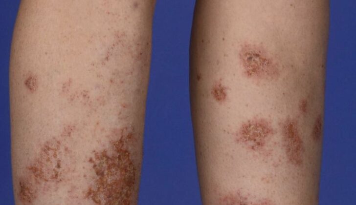
Acute skin lesions may initially appear as small bumps or blisters that merge together to form raised patches. As these lesions grow, they become symmetrical, clearly outlined, and appear in round or coin-shaped patches of red skin. These patches, or plaques, can vary in size from as small as 1 cm to as large as 10 cm. As the lesions progress, they may turn into drier scaled patches and might thicken over time. The intensity of itchiness linking to these lesions could range from mild to severe.
Activities that reduce the skin’s natural moisture, such as using harsh soaps or taking hot and lengthy showers, can cause the lesions and their symptoms to get worse. Areas of the body most frequently affected include the lower and upper limbs, followed by the trunk. The face and scalp are usually unaffected. After the lesions have healed, there is often a change in skin color in the affected area.
Under close skin examination, the scales may show shiny yellow clumps and irregularly distributed reddish-brown globules.
Testing for Nummular Dermatitis (Nummular Eczema)
Nummular dermatitis, also known as discoid eczema, is a skin condition recognized by its typical symptoms of red, itchy, coin-shaped rashes on the skin, usually found in individuals with generally dry skin. This diagnosis is most often made based on these classic symptoms.
To aid in the diagnosis, a doctor may perform a dermoscopy, which is a procedure that uses a specialist microscope to examine the skin in detail. Generally, additional lab tests are not usually needed. However, in certain situations, the doctor may use extra tests to differentiate it from other conditions that cause similar round, red skin patches.
Such additional tests could include a potassium hydroxide examination, which involves taking a scraping of the skin and checking it under a microscope for fungal infections. If the rashes are crusted or oozing, a skin swab might be taken and sent for bacterial culture to check for an infection. Biopsy, which involves removing a small piece of the affected skin for further examination, could also be performed.
If the skin condition doesn’t improve with treatment or if there’s a history indicating possibility of allergic contact dermatitis (a type of skin inflammation caused by contact with an allergen), then patch testing might be performed. In patch testing, small amounts of potential allergens are applied on the skin using patches to see if there’s a reaction.
Treatment Options for Nummular Dermatitis (Nummular Eczema)
Managing nummular dermatitis, a type of skin condition, mainly involves taking care of your skin, keeping it hydrated, and avoiding things that irritate it.
For general care, it’s suggested to moisturize your skin often with thick products like petroleum jelly. Some moisturizers that contain ceramides can also help keep your skin hydrated. It’s advisable to take short showers of less than 5 minutes with lukewarm water, using gentle soaps that don’t dry out your skin. Apply moisturizer right after your shower when your skin is still a bit damp. Wearing comfortable, soft clothing, avoiding scratchy fabrics, and using a humidifier can also help keep your skin from drying out.
When it comes to creams and ointments, strong corticosteroids can be applied to the affected skin once or twice a day to help reduce inflammation and itchiness. Calcineurin inhibitors (like tacrolimus, pimecrolimus) can be used as an alternative to corticosteroids. An injection of a specific medicine called triamcinolone can be considered for stubborn spots.
If the condition is widespread and creams aren’t enough, you might be recommended a type of treatment called narrowband UVB light therapy, also known as phototherapy. However, it’s important to understand that this therapy should be used with caution as it might increase your risk of skin cancer.
If light therapy is not an option, there are other treatment methods that can be used involving medicines that modify or suppress the immune system, like systemic (or overall) corticosteroids, methotrexate, cyclosporine, and dupilumab. These treatments are usually considered for severe cases when other treatments haven’t worked.
If there’s a risk that your skin condition has been infected by bacteria, your doctor might prescribe you antibiotics, either topical (applied to the skin) or oral, depending on the extent of the infection. Antihistamines, which are medicines commonly used to treat allergies, can also be used to relieve severe itchiness, especially at night.
What else can Nummular Dermatitis (Nummular Eczema) be?
When a doctor is diagnosing nummular eczema, they must consider that it could be a number of other skin conditions. Here’s a list of possible alternatives and their characteristics for easy understanding:
- Atopic dermatitis: This condition also looks like nummular eczema, but it needs certain criteria for diagnosis.
- Allergic contact dermatitis: This one also could show up with nummular lesions. Through patch testing, what is causing the allergy can be identified.
- Plaque psoriasis: Usually shows up as clear, red plaques with silver scales, most commonly on scalp, elbows, or knees, although they can be anywhere.
- Stasis dermatitis: Usually connected with problems like edema, varicose veins and atrophic scars. The ankle and lower leg can show red patches that may turn into scaly plaques.
- Asteatotic eczema: Also called “eczema craquelé”, it shows up as redness and small scales on both lower legs.
- Lichen aureus: This one will show small red to brown spots or plaques, usually on lower legs, but occasionally on the trunk and arms.
- Fixed drug eruption: Shows up as round, red to brown patches or swollen plaques that reappear in the same place when the patient is exposed to the implicated drug.
- Erythema annulare centrifugum: Characterized by round or arc shaped plaques with central clearing and a scale at the outer edge.
- Bullous pemphigoid: This may first appear as itch-inducing, eczema-like lesions for a long time before classic blisters show up.
- Tinea corporis: Appears as one or many circular, red scaly plaques with central clearing. Confirming diagnosis can be done by looking for characteristic features in a solution of potassium hydroxide.
- Majocchi granuloma: This is characterized by multiple pimple-like spots that come together to form reddish scaly plaques.
- Impetigo: Shows up as “honey-colored” or golden-crusted spots and plaques with a red base.
- Secondary syphilis: Shows up as widespread pink, red, purple, or brown spots and plaques with an overlying scale. The palms and soles are often affected.
- Pityriasis rosea: Presents as pale pink circular or oval patches or thin plaques with a fine white ring of scale.
- Patch or plaque stage mycosis fungoides: This skin condition demonstrates as reddish to slightly colored-up patches or thin plaques with overlying fine scale. The affected areas are usually those protected from sunlight such as the buttocks.
- Squamous cell carcinoma in situ: This skin condition typically manifests as a single (or rarely multiple) red, scaly, well-defined, thin patch with a circular or irregular shape.
This information emphasizes the importance of a thorough examination by a healthcare provider to correctly diagnose skin conditions.
What to expect with Nummular Dermatitis (Nummular Eczema)
Nummular dermatitis is a skin condition that typically follows a long-term pattern. This means that its symptoms can come and go over a span of months or even years. It’s common for the condition to improve for some time, only to have symptoms reappear later. This cycle is also known as relapses and remissions.
Possible Complications When Diagnosed with Nummular Dermatitis (Nummular Eczema)
The damaged skin barrier can lead to further infection in the affected area, most commonly caused by a type of bacteria called Staphylococcus aureus. Infected skin lesions may produce a pus-like discharge and develop a thick, golden crust that’s more noticeable than non-infected lesions.
To identify the bacteria and determine its sensitivity to different antibiotics, a swab of the infected skin can be taken and sent for laboratory testing. Based on the prevalence of antibiotic-resistant bacteria in your local area, the doctor may initially prescribe doxycycline or a different antibiotic effective against Staphylococcus aureus. The treatment plan can then be adjusted based on the lab results.
As with any inflammation of the skin, some discoloration may occur after the condition improves. This might include redness, or a lighter or darker patch of skin.
Potential Consequences of Infected Skin Lesions:
- Further infection due to a damaged skin barrier
- Infections most frequently caused by Staphylococcus aureus
- Pus-like discharge from affected skin
- Thick, golden crust developing on lesions
- Skin discoloration following inflammation
- Post-infection redness or hypo- or hyperpigmentation.
Preventing Nummular Dermatitis (Nummular Eczema)
Doctors often advise that changing certain behaviors can greatly help improve a skin condition known as nummular dermatitis. This includes taking short showers that are less than or equal to 5 minutes in duration, and using lukewarm water instead of hot water.
Apart from this, it’s important to use gentle hydrating liquid cleansers during showers. After showering, while your skin is still wet, apply a moisturizer or ointment – something simple like petroleum jelly can work well. Remember, the key is to apply it immediately after your shower when your skin is still damp.
Patients are also encouraged to avoid wearing tight clothing or using itchy fabrics such as wool as these can irritate the skin further. Finally, it’s crucial that patients follow the medication schedule and instructions provided by their doctor.


