What is Osteoma Cutis?
Osteoma cutis, or cutaneous ossification, is a rare, harmless skin disease where bone forms in the skin or just beneath it. The disease can occur on its own, known as primary osteoma cutis, or can develop alongside an existing condition like an injury or an inflammatory or cancerous disease, known as secondary osteoma cutis. Both children and adults can be affected by this condition. Primary osteoma cutis is less common, accounting for only 15% of cases, while secondary osteoma cutis makes up the remaining 85%. The exact cause of this skin condition is not fully understood yet.
What Causes Osteoma Cutis?
Osteoma cutis is a condition where bone forms within the skin. This can occur in two ways: primary or secondary. If it is “primary”, it develops without any previous skin condition. It can happen alone or along with a condition known as metabolic syndrome, which is a cluster of conditions including high blood pressure, high blood sugar, excess body fat around the waist, and abnormal cholesterol levels.
If it’s considered “secondary”, then it’s associated with conditions that cause inflammation, scars, birth defects, or abnormal growths in the skin.
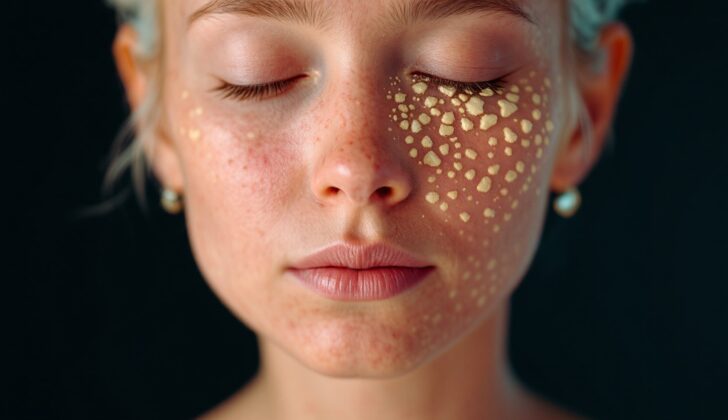
Some scientists believe there is a link between this condition and long-lasting acne. They suggest that roughly 85% of osteoma cutis cases occur as a result of persistent acne. This is often noticed in particular areas, such as the face for females, and the scalp or chest for males.
Another recent discovery has linked osteoma cutis to certain gene mutations, specifically in the GNAS1 gene. This gene is important for regulating a couple of rare genetic bone disorders known as progressive osseous heteroplasia and Albright hereditary osteodystrophy.
Risk Factors and Frequency for Osteoma Cutis
Osteoma cutis is a condition that leads to bone formation in the skin and it represents 14% of all skin ossifications. This can occur in both adults and children, but it’s more common in females. These unusual bone formations on the skin usually start appearing in one’s 20s or 30s. This condition can either start to develop on its own (primary osteoma cutis) or as a result of inflammation or the growth of abnormal masses of tissue (secondary osteoma cutis).
- Osteoma cutis is responsible for 14% of skin ossifications.
- It can affect adults and children but is more common in females.
- These bone formations typically start showing up around a person’s 20s or 30s.
- Osteoma cutis can either start developing on its own or as a result of inflammation or abnormal tissue growth.
- The most common places for these formations are the face in females and the scalp in males.
- Other areas where these bone formations can occur include the breasts, buttocks, and extremities.
- Though rare, osteoma cutis can also develop in the mucosae such as the tongue.
Signs and Symptoms of Osteoma Cutis
Osteoma cutis is a condition where bone forms within the skin. The appearance of this condition can vary greatly. It can show up as one or multiple areas and can be anywhere from almost invisible (0.1 cm) to quite noticeable (5.0 cm). These areas may appear as raised bumps or nodules, flat patches, or as smaller, grain-like lesions. When you touch them, they feel hard and can sometimes cause the skin to turn white or yellowish.
In rare cases, the skin over the bony area might break open, releasing tiny bone fragments. This unique presentation is known as perforating osteoma cutis.
There are four specific types of osteoma cutis:
- Solitary: Just one nodule that can appear anywhere on the skin.
- Widespread: Multiple bony nodules appearing all over the skin, usually present from birth.
- Plate-like: Appears as a bony plate, often noticed at birth or within the first few years of life.
- Multiple miliary osteomas: Features multiple, small pinpoint bone formations within the skin.
Doctors diagnose plate-like osteoma cutis by confirming the following things:
- The patient has had a bony plate since birth.
- The patient does not have a metabolic disorder.
- There may or may not be osteomas in other parts of the body.
- There have been no incidents of trauma, infection, or other significant events.
Medical professionals would also check for any unusual features in patients with osteoma cutis. This condition sometimes goes hand in hand with diseases like progressive osseous heteroplasia, Albright’s hereditary osteodystrophy, or fibrodysplasia of progressive ossification.
Progressive osseous heteroplasia is a rare condition that can be inherited. It causes progressive bone formation within the skin during infancy, and as the child grows, deeper tissues also turn to bone. This condition doesn’t come with hormonal abnormalities, but it can slow down considerably in adulthood. However, it can cause joints to stiffen and limbs to grow slower than usual.
Albright’s hereditary osteodystrophy presents with short height, a round face, obesity, and intellectual disabilities. Between 25 to 50 percent of individuals with Albright’s hereditary osteodystrophy could also have osteoma cutis. This condition often comes with hormonal abnormalities as well.
Testing for Osteoma Cutis
Your doctor might carry out blood tests to check your calcium and parathyroid hormone levels if Albright’s hereditary osteodystrophy – a rare genetic disorder affecting bone development – is suspected.
Also, certain imaging tests like traditional X-rays, Computerized Tomography (CT) scans, or skin ultrasound could be used to examine any hardening or calcification in your soft body tissues.
Conventional 2D X-rays can be challenging to interpret, particularly when it comes to pinpointing the precise location of osteoma cutis, a condition in which bone forms in the skin.
On the other hand, the Cone-beam CT scan – which produces three-dimensional (3D) images – can greatly help with detecting and diagnosing osteoma cutis. Osteoma cutis appears as a regular, often circular shaped lesion on these scans and can sometimes show a less dense center. It has the same degree of solidity as bone and can sometimes have different shapes, like a donut, snowflake or even washer.
A skin biopsy, which involves taking a small sample of skin for laboratory analysis, can certify a diagnosis of osteoma cutis.
Treatment Options for Osteoma Cutis
When it comes to treating osteoma cutis, a condition wherein small bony deposits form in the skin, various factors are considered. These factors include how severe the condition is, how much it has spread, where it is located on the body, and what caused it.
Several treatment options can be chosen from, ranging from non-invasive to invasive methods.
Non-invasive treatments are simpler and do not require any kind of surgery. They include the usage of a tretinoin cream, which is a topical medication often used for skin conditions. However, this treatment seems to best work for smaller and shallower lesions, or damaged areas in the skin.
On the other hand, invasive treatment options call for a more direct approach to remove these bony deposits. They include a combination of dermabrasion (a technique that exfoliates the outermost layers of the skin) and punch biopsy (a procedure to obtain skin samples), various forms of laser treatments like the Erbium: YAG laser, and surgical methods like scalpel incisions and curettage (scraping of skin). Among laser procedures, CO2 and erbium: YAG lasers can cause changes in skin color. However, there have been successful cases where the Q-switched neodymium: YAG laser treatments didn’t lead to any color changes.
In cases of secondary osteoma cutis, which develops as a result of another disease or condition, it’s essential to also investigate and treat any underlying issues. For example, if osteoma cutis is linked with pseudohypoparathyroidism (a condition that causes low levels of calcium in the blood), then patients may also require therapy involving calcitriol (a form of vitamin D) and calcium to manage low calcium levels.
What else can Osteoma Cutis be?
The medical process to determine the exact diagnosis involves considering a range of other possible conditions. This is known as differential diagnosis. For osteoma cutis, the following conditions are considered as potential diagnoses based on the examination of tissue under a microscope (histological differential diagnosis):
- Pilomatrixoma in its ossified (hardened) form
- Osteochondroma (a type of bone growth)
- Ossified hair follicle, for instance within a melanocytic nevus (type of mole)
- Osteosarcoma (bone cancer) in its extraskeletal format found in the skin or subcutaneous tissue (beneath the skin)
- Cutaneous calcinosis (calcium deposits in the skin)
And based on medical imaging like X-Ray or CT scan (radiological differential diagnosis), the following conditions might also be considered by the physicians:
- Calcified phleboliths (small calcified blood clots)
- Calcified nodules due to surgical clips, wires, or sutures that may look similar to osteoma cutis on imaging
What to expect with Osteoma Cutis
Osteoma cutis is a type of harmless tumor that does not spread to other areas or disrupt surrounding structures. The outlook for this condition is excellent. This means that the long-term health of individuals with this condition is usually very positive.
Possible Complications When Diagnosed with Osteoma Cutis
Laser treatments that use CO2 and erbium:YAG lasers may cause changes in the color of the skin.
Preventing Osteoma Cutis
If you notice any hard lumps on your skin, it’s recommended to see a skin doctor (a dermatologist). They can provide information and support about a harmless skin condition called osteoma cutis, including why it happens, what to expect, and how it can be treated. Your healthcare provider may share useful website resources to help you better understand osteoma cutis.
Education and understanding are key in preventing health issues that may lead to osteoma cutis. Learning about the importance of keeping up with regular healthcare appointments and understanding the treatments you’re receiving can be very valuable. This will ensure that any changes in your condition are monitored closely, and any necessary adjustments to your treatment can be made promptly.


