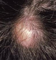
What is Pilar Cyst?
Pilar or Trichilemmal cysts are common skin issues that you may not realize because they occur in less than 10% of people. These cysts are the most common type of skin cyst and usually affect the scalp. The good news is that these cysts don’t develop into harmful cancerous growths. They tend to show up randomly and are filled with a protein called keratin. The walls of these cysts are made up of the same material that you’d see in the outer part of a hair follicle.
Now, there’s something called a proliferating trichilemmal cyst, which is basically a tumor form of a Pilar cyst. This is even less common, appearing in less than 3% of all pilar cyst cases. This type of cyst can break open (ulcerate) and might be more aggressive, but it’s important to remember that this is rare.
What Causes Pilar Cyst?
Trichilemmal cysts, also known as pilar cysts, can sometimes be inherited from parents – this is known as an autosomal dominant trait. People with this inherited type of pilar cysts generally start experiencing them at a younger age and may have multiple cysts appear at the same time. These cysts start to form in the layer of skin between the oil-producing gland (the sebaceous gland) and the tiny muscle at the base of a hair (the arrector pili muscle).
Their most common location is on the head, especially the scalp. These cysts typically grow very slowly – it can take several years for them to get big.
Risk Factors and Frequency for Pilar Cyst
Trichilemmal cysts, also known as pilar cysts, tend to occur more frequently in young people. They don’t favor any particular race and are found more often in women compared to men. It’s possible for these cysts to run in families as in some instances, they are inherited in an autosomal dominant manner.
Signs and Symptoms of Pilar Cyst
Pilar cysts, also known as trichilemmal cysts, are usually diagnosed based on their appearance and symptoms. More often than not, a person tends to have more than one cyst, though sometimes only a single cyst might appear. Since these cysts develop from hair follicles, they are most commonly found on areas with a lot of hair. This means they are primarily found on the scalp, though they can also appear on the face, head, and neck.
Most of the time, pilar cysts don’t cause any problems unless they harden or break open, which can lead to inflammation and pain in the area of the cyst. Sometimes, if a cyst forms on a pressure point or bony area, it can also cause discomfort. Pilar cysts typically appear to be smooth, movable, firm lumps that are the same color as the person’s skin. They are usually distinct and clearly separate from the surrounding tissue.
It is also worth mentioning that a family history of pilar cysts can be significant. This is because this condition can be inherited in what is known as an autosomal dominant pattern. This means that if one of your parents has the condition, you have a 50% chance of getting it too. Generally, pilar cysts grow slowly but occasionally they might grow rapidly. A sudden increase in size might suggest an infection or even a change into a cancerous growth.
While pilar cysts can affect anyone, they seem to be more common in young females as compared to males.
Testing for Pilar Cyst
Pilar cysts are typically diagnosed through a check-up, where the doctor evaluates your signs and symptoms. Usually, no extra tests are needed. However, sometimes a doctor might need to use imaging tools like x-rays or scans to rule out any other possible conditions. These tools are especially helpful if the cyst is found in the middle of your head or neck because they can help measure the size of the cyst and check if it’s affecting the brain and spinal cord.
For instance, if your doctor wants to see if the cyst is affecting the skull (the bone that encloses your brain), a CT scan (a type of x-ray that gives detailed pictures of your insides) is the best option. On the other hand, if your doctor wants to look at soft tissues (such as muscles, ligaments and organs) or small invasions deep inside your body, an MRI scan (a type of scan that uses strong magnetic fields and radio waves to produce detailed images of the inside of the body) might be used.
Treatment Options for Pilar Cyst
The main form of treatment for this is to entirely remove the affected area through surgery, including the walls of the cysts. This helps to reduce the chance that the cysts might return. After the cysts have been removed, they are sent to the pathology department for testing to confirm the diagnosis.
The preferred treatment is the complete surgical removal of the cyst itself. However, if the cysts return or become inflamed (swollen and painful), it’s usually not advisable to try and remove them surgically. Instead, it’s best to wait until the swelling goes down before considering surgery.
If a cyst becomes inflamed, a sample, or ‘swab’, is taken from the wound, and tested (‘culture sensitivity’) to see if there is an infection present. This can help decide the best course of treatment.
Proliferating trichilemmal cysts, a specific type of cyst that can multiply rapidly, may need additional treatment beyond just surgery. These could include multiple rounds of surgery, radiation therapy, and/or chemotherapy. Radiation therapy uses high-energy beams to kill cells, and chemotherapy uses drugs to kill cells or stop them dividing and multiplying.
What else can Pilar Cyst be?
These are some skin conditions that may be confused with others due to their somewhat similar appearances:
- Acne keloidalis nuchae
- Cutaneous lipomas
- Dermoid cyst
- Epidermal inclusion cyst
- Favre-Racouchot syndrome (also known as nodular elastosis with cysts and comedones)
- Pilomatrixoma
- Steatocystoma multiplex
What to expect with Pilar Cyst
Overall, pilar cysts, which are fluid-filled lumps under the skin, usually have a positive outcome, even if there are complications. Possible side effects of these cysts include inflammation, infection, and in rare cases, they can become cancerous. It’s important for the patient and their family to receive counseling as this condition can be passed down from parent to child (a phenomenon known as autosomal dominant inheritance).
Possible Complications When Diagnosed with Pilar Cyst
A pilar cyst can lead to discomfort, particularly in areas subjected to pressure. Additional problems can occur such as swelling, changes in appearance, infection, and hardening of the cyst due to calcium build-up. If the cyst is surgically removed, complications may involve bleeding, pain, infection, and formation of scar tissue.
Potential Complications:
- Discomfort, especially in pressure areas
- Swelling
- Changes in appearance
- Infection
- Hardening of the cyst due to calcium build-up
- Bleeding after surgical removal
- Pain after surgery
- Infection post-surgery
- Formation of scar tissue post-surgery
Recovery from Pilar Cyst
If you’ve had a Pilar cyst (a small, non-cancerous lump under the skin) removed through surgery, it’s crucial that you keep the surgical area clean. This involves changing the dressing daily and cleaning the area with normal saline (a type of salt water solution) and a disinfectant. It’s also advised that the stitches should be covered with a gauze dressing, especially in the first few days after surgery. Generally, your doctor will want to remove the stitches about 7 to 10 days afterwards, depending on where the cyst was and how well the wound is healing.


