What is Porokeratosis?
Porokeratosis is a rare skin condition that results in rough, raised bumps or ring-shaped patches with a border that sticks out. This happens because of unusual skin cell growth. Under a microscope, the developed skin cells form a unique column-like structure which doctors refer to as ‘cornoid lamella’. Different types of porokeratosis exist, including:
* Disseminated superficial actinic porokeratosis
* Classical porokeratosis of Mibelli
* Porokeratosis plantaris, palmaris, et disseminate
* Linear porokeratosis
There’s also some less common types like:
* Genitogluteal porokeratosis
* Facial porokeratosis
* Giant porokeratosis
* Porokeratosis ptychotropica
* Hypertrophic verrucous porokeratosis
* Eruptive pruritic papular porokeratosis
* Follicular porokeratosis
* Reticulate porokeratosis
Although it’s possible for a person to have more than one type of porokeratosis, it’s quite uncommon. It’s worth noting that porokeratosis can potentially evolve into skin cancer. To check for porokeratosis, doctors usually take a sample from the raised border of the affected skin. Even though there are multiple treatment options available — including creams, medications and surgery — there’s no one-size-fits-all treatment guideline for this condition.
What Causes Porokeratosis?
Porokeratosis is a skin condition that has been known about for over 100 years, but the exact causes and nature of the disease are still not completely understood. It gets complicated because there are a variety of factors that might cause it, including exposure to ultraviolet (UV) light, frequent skin friction or trauma, genetics, infections, cancer, and having a weakened immune system like in transplant patients.
There are six different types of porokeratosis, which include: disseminated superficial porokeratosis (DSP), disseminated superficial actinic porokeratosis (DSAP), eruptive disseminated porokeratosis (EDP), porokeratosis of Mibelli, linear porokeratosis, and punctate palmoplantar porokeratosis. The most common type is disseminated superficial actinic porokeratosis.
One detail that has been learned about DSAP is that it can be inherited in a pattern where one parent carries the gene and has the chance to pass it onto their child. A key gene that has been found to contribute to this skin condition plays a critical role in the production of cholesterol and protection of the skin from UV light. This finding has been important for developing medications to treat the disease.
There’s also a confirmed link between exposure to UV light and the development of porokeratosis. Some experiments have even succeeded in creating cases of DSAP by exposing subjects to UV light.
Certain drugs and types of treatment might also be linked to the development of porokeratosis. A review of patient records found that biological usage, phototherapy, and radiotherapy were common treatments used by patients who developed the skin condition.
It’s also worth noting that cases of porokeratosis of Mibelli and DSAP are often found in patients who have undergone some kind of therapy that weakens the immune system. In fact, a study found that more than 10% of organ transplant patients who were on this kind of therapy developed porokeratosis.
Though there is still much to learn about this condition, there is evidence that it can develop at any time from three weeks to 14 years after starting a therapy that weakens the immune system.
Risk Factors and Frequency for Porokeratosis
Porokeratosis is a rare skin condition that can appear in different forms and on different parts of the body. It primarily appears on sun-exposed areas of the body, but it can be seen anywhere including the limbs, palms, and soles of feet. The condition typically shows up around the age of 50, but it can affect anyone, regardless of their sex.
Although it’s primarily seen in adults, cases have also been reported in young children. Sun exposure seems to increase the risk of a particular variant of the disease, pointing to UV light as a possible risk factor.
Porokeratosis is often related to immune suppression, often seen in patients who have had organ transplants, are dealing with inflammation, or have cancer. It has the potential to develop into a non-melanoma skin cancer in 6.9% to 30% of cases, often squamous cell carcinoma and at times basal cell carcinoma.
- A study of 281 patients with porokeratosis unveiled various risk percentages for malignant transformation based on the type of porokeratosis:
- Linear porokeratosis: 19%
- Porokeratosis plantaris, palmaris, et disseminate: 10%
- Porokeratosis of Mibelli of any size: 8%
- Disseminated superficial actinic porokeratosis: 3%
It’s important to note that porokeratosis lesions do not immediately become cancerous – on average, it takes about 36 years for a lesion to become cancerous. Even though porokeratosis is usually acquired, there often seems to be a family history, implying a potential genetic component to the disease.
Signs and Symptoms of Porokeratosis
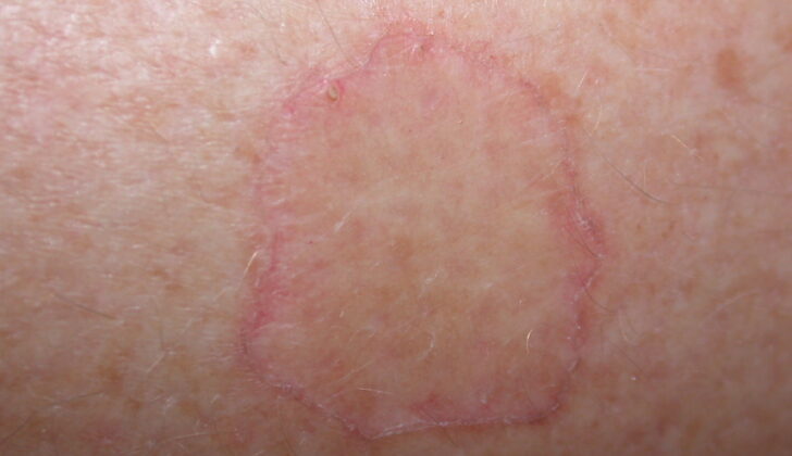
Porokeratosis is a skin condition that shows up as rough bumps or ring-shaped plaques that expand outwardly with a raised rough border. The middle part of these spots can seem a bit sunken. The spots might be itchy, and sometimes they can be there for several years before being diagnosed. There is also a form of porokeratosis that emerges as small pink to brown raised spots.
Porokeratosis mostly develops on the arms and legs or the trunk, but it can also appear on the face, the urinary and genital regions, and the scrotum. It’s important to remember that porokeratosis can have a variety of manifestations, including large destructive lesions and involvement of scars left by burns. High attention should be given to relatively large spots of porokeratosis situated on parts of the body exposed to the sun, particularly on the arms and legs, and those that have been there for a long time since it increases the risk of turning malignant.
Testing for Porokeratosis
For the evaluation of porokeratosis, which is a rare skin condition, the best approach is to perform a biopsy, a medical test involving the removal of cells or tissues for examination. The biopsy is specifically taken from the raised edges of the affected skin. This process will show a structure called the cornoid lamella, which confirms the presence of porokeratosis.
Another useful tool for diagnosis is called dermoscopy, a test that uses a special magnified lens to examine the skin. When porokeratosis is present, this skin examination might show a characteristic pattern of central brown pigmentation with blue-gray dots, surrounded by a single pale-colored band with a white streak at the outer part.
Even though porokeratosis is relatively rare, particularly certain types like genitourinary porokeratosis that affect the genital and urinary tract regions, it’s important to be aware of this condition due to the potential risk of it transforming into skin cancer.
Treatment Options for Porokeratosis
Porokeratosis is a term that refers to a variety of skin conditions. Treatment options for these conditions can vary depending on the specific type of porokeratosis a person has, and can include creams, medications, or even surgical procedures. However, it’s important to note that there isn’t one international standard for treating porokeratosis due to the lack of extensive research.
There are different types of porokeratosis and they can be treated as follows:
– Classical porokeratosis of Mibelli is often successfully treated with a cream called imiquimod.
– A type of porokeratosis called DSAP could be treated with Diclofenac 3% gel. This gel has been found to be reasonably effective and generally causes few side effects. Radiotherapy, which uses radiation to kill cells, has also been used with around 50% effectiveness, but it can cause side effects such as changes in skin color, hair loss, skin thinning, small blood vessel appearance, fibrosis (formation of excess fibrous tissue), and even secondary skin malignancies. Some forms of vitamin D3 have been proven effective as well as certain types of laser therapy and cryotherapy, which uses cold treatment. A 2023 study suggested a 2% levostatin cream as a primary treatment due to excellent results.
– Linear porokeratosis often responds well to topical or oral retinoid medications, which are derived from vitamin A. DSP, another type of porokeratosis, is treated similarly to DSAP, excluding the use of topical statin. Other medications, including dimethyl sulphoxide, oral tofacitinib and a traditional Chinese herbal medicine called Huang-Lian-Jie-Tang, have been tested and shown to provide relief of itching in small studies.
– Eruptive Disseminated Porokeratosis is a subtype of DSP that tends to go away on its own within a month.
– Punctate porokeratosis, another variation, has a less clear treatment approach due to limited data, but oral retinoid medications such as isotretinoin, etrenitate, and acitretin have been documented to be the most effective.
Surgical treatments, like sharp excision, curettage (scraping of the skin) and electrodesiccation (destroying tissue with heat), may be used in areas where creams or medications are not practical or approved for use.
In general, patients with only a few small skin lesions and who are okay with potential aesthetic changes may opt for surgical or laser procedures. Those with numerous, large lesions are usually better suited for topical medication therapies. Lastly, if a patient does not want to undergo surgery or intensive topical medication therapy, topical keratolytic agents, which help to break down the outer layer of skin, may be a more suitable option.
What else can Porokeratosis be?
Porokeratosis is a skin condition that can look like various other skin problems. These include:
- Psoriasis
- Lichen simplex chronicus (chronic skin inflammation)
- Hypertrophic lichen planus (skin disorder causing bumps)
- Skin tuberculosis
- Bowen disease (skin cancer)
- Candidiasis (yeast infection)
- Reactions to irritants
- Allergic contact dermatitis (skin inflammation caused by contact with an allergen)
What to expect with Porokeratosis
Porokeratosis is a skin condition that doesn’t go away on its own. It’s very rare for these skin changes to disappear without any medical or surgical treatment. Porokeratosis is a condition that could potentially turn into skin cancer. All types of porokeratosis have a chance of becoming nonmelanoma skin cancer, ranging from a 6.9% to 30% chance.
Most often, porokeratosis turns into a type of skin cancer called squamous cell carcinoma. It less commonly changes into basal cell carcinoma. If the porokeratosis has not yet turned into cancer, getting rid of it through surgery can provide a complete cure.
Two specific types of porokeratosis, called linear porokeratosis and giant porokeratosis, are at a higher risk of becoming malignant, or cancerous. On the other hand, cancer rarely develops from a form of porokeratosis called disseminated superficial actinic porokeratosis.
Possible Complications When Diagnosed with Porokeratosis
The complications related to porokeratosis mostly depend on the kind of treatment chosen. If a surgical operation is chosen, it can lead to issues such as infections, bleeding, and the formation of scars. Applying topical treatments could potentially irritate the skin, lead to skin thinning, cause itching or dryness, or even result in changes in skin pigmentation.
If porokeratosis is not diagnosed early or treated adequately, it might also lead to other issues. One such serious problem could be the conversion of the condition into a malignancy.
Possible side effects:
- Infections due to surgical interventions
- Bleeding post surgery
- Scarring as a result of surgical procedures
- Aggravated skin irritation due to topical treatments
- Skin thinning with topical application
- Itching or dry skin generated by topical treatments
- Changes in skin pigmentation
- Possibility of the development of malignant tumors
Preventing Porokeratosis
Porokeratosis is a type of skin condition that has the potential to turn into skin cancer. This condition is characterized by small, hardened bumps (also known as keratotic papules) or flat, round spots (referred to as annular plaques) with raised edges on the skin. Research shows that these spots can become cancerous in about 7% to 30% of cases, most commonly turning into a type of skin cancer called squamous cell carcinoma.
Often, porokeratosis is seen in areas of the skin that get a lot of sun and usually becomes apparent when a person is in their 50s. Interestingly, there are several different types of porokeratosis, but researchers are still trying to determine the root cause and how these skin spots develop. The most severe types of porokeratosis are the linear and giant forms, as they have a higher chance of turning into cancer. In contrast, a type known as disseminated superficial actinic porokeratosis is less likely to become cancerous. The causes of porokeratosis could be many, including exposure to ultraviolet (UV) light, skin injuries, genetic factors, infections, and conditions that weaken the immune system.
To make a concrete diagnosis, doctors often need to take a sample (biopsy) from the raised edge of the skin spot. This biopsy can identify a distinctive feature called the cornoid lamella, which confirms porokeratosis. This is important, as porokeratosis can look like other skin conditions such as psoriasis, lichen simplex chronicus, hypertrophic lichen planus, skin tuberculosis, Bowen disease, candidiasis, irritants, and allergic contact dermatitis.
There are several ways to treat porokeratosis, including creams, medications, and surgery. It is recommended for patients to have yearly check-ups to watch for signs of the condition turning into cancer and to lower this risk. Doctors will also check for any underlying conditions that might weaken the immune system and will provide advice on sun protection habits.
If a patient’s porokeratosis spots are large, numerous, or have been present for a long time, these are considered risk factors for the condition becoming cancerous. In these cases, check-ups should be scheduled twice a year. If a spot begins to show signs of turning into cancer, it is important to see a doctor right away for possible removal or further biopsy.


