What is Premalignant Fibroepithelial Tumor of Pinkus?
In 1953, a doctor named Herman Pinkus first discovered a type of pre-cancerous skin growth known as the Fibroepithelial Tumor of Pinkus (FEP). There has been some disagreement over how to classify this type of tumor, but it has a distinctive appearance that sets it apart from other similar skin growths. There are several key ways to tell the difference between FEP and another type of skin cancer known as basal cell carcinoma (BCC).
One major difference is that FEP contains specific cells known as Merkel cells, while BCC does not. FEP also tends to occur more frequently in women and is not related to sun exposure. It most commonly appears on the lower body. BCC, however, usually appears on parts of the body that get a lot of sun exposure.
Another important thing to note is that FEP is usually not aggressive; there have been no reported cases of it invading other tissues or spreading to other parts of the body. On the other hand, BCC can invade surrounding tissues and spread if it’s not treated in time.
What Causes Premalignant Fibroepithelial Tumor of Pinkus?
The beginning of FEP, or Fibroepithelioma of Pinkus, is not well understood. Some experts suggest that FEP may develop from a common skin growth known as seborrheic keratosis. Others propose that FEP could be an extension of a type of skin cancer called basal cell carcinoma, spreading through sweat glands. This is based on findings from microscope studies of FEP, which show some structures similar to sweat glands among the collection of cells that make up FEP.
However, this idea is disputed by critics who argue that sweat glands grow straight down and do not merge with each other often, making such sideways spread unlikely.
Other research hints that the thin, connecting threads that are a key part of FEP’s structure are PHLDA1 positive, meaning they contain a particular protein which could signal a kind of skin cell growth or “hyperplasia” where these cells are excessively forming. This is where the FEP’s characteristic appearance comes from. These threads also contain a large number of Merkel cells, a type of cell involved with the sense of touch.
The clusters of basaloid nubbins – that is, small rounded lumps of cells with similarities to the base layer of the skin – found within FEP are suggested to potentially develop into nodular BCC, a form of basal cell carcinoma. This is particularly noteworthy as it’s not unusual to observe nodular BCC near or connected with FEP. Moreover, these basaloid nubbins test positive for nestin, a protein usually identified in the support tissue of basal cell carcinoma.
Risk Factors and Frequency for Premalignant Fibroepithelial Tumor of Pinkus
Fibroepithelial polyps (FEP) are considered uncommon. However, some believe that these types of tumors might not be as rare as we think – they might just be misdiagnosed often. FEP tends to show up in men between the ages of 40 and 60, but they can appear at any age, even in children.
Signs and Symptoms of Premalignant Fibroepithelial Tumor of Pinkus
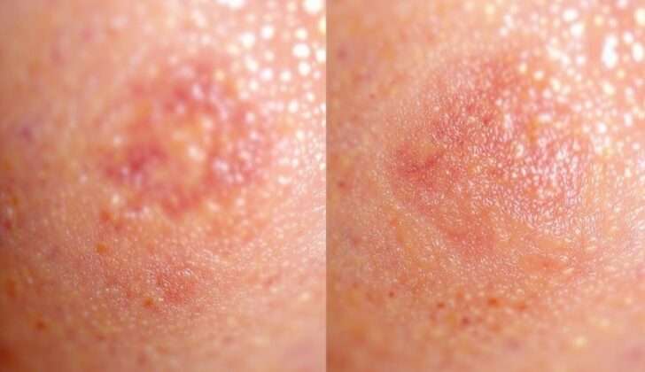
Fibroepithelial polyp (FEP) usually appears as one or more growths on the skin. These growths can look like little bumps or stalk-like protrusions. Depending on the type, the color of the tumor can vary – it might be pink, tan, brown, or even the same color as the skin. We can categorize FEP into two types based on its color: pigmented (colored) and non-pigmented (skin-colored). Sometimes, there can be scratches or minor damage on the surface of the growths, but they don’t usually have deep sores. Interestingly, these growths typically occur on the trunk of the body and are not related to exposure to ultraviolet (UV) light.
Testing for Premalignant Fibroepithelial Tumor of Pinkus
Some experts recommend using skin surface microscopy, also known as dermoscopy, and a special type of microscope called reflectance confocal microscopy (RCM) to help diagnose certain skin conditions. Using dermoscopy, they may see small branching blood vessels and tiny spots around the edges. When using a specific type of light (polarized light), they could see short, white streaks, known as crystalline or chrysalis structures. If the area of the skin being looked at has color to it, the dermoscopy could display gray-brown areas and/or gray-blue dots, which suggests a specific skin condition called Fibroepithelioma of Pinkus (FEP). In one study, a researcher was able to accurately diagnose FEP in nine out of ten patients using dermoscopy.
When it comes to the reflectance confocal microscopy, this device can show the unique ‘fenestrated pattern’ or a pattern with small openings at the layer where the skin and the underlying tissue meet. This pattern shows ‘holes’ of fibrous tissue surrounded by nice rows of cells. This pattern is a well-recognised sign of these types of tumors.
An analysis of the tissue under a microscope, after it has been surgically removed, can help to confirm the diagnosis.
Treatment Options for Premalignant Fibroepithelial Tumor of Pinkus
The treatment for a fibroepithelial tumor of Pinkus, a type of skin tumor, involves identifying the tumor and then completely removing it through surgery. This condition might be overlooked and under-reported because it often appears harmless on the surface.
There are different ways to remove this type of tumor. One method includes using electricity to dry out the tumor (electrodesiccation) and then scraping it off (curettage). However, because this tumor is rare and often overlooked, there’s not enough evidence to say how effective these methods are.
Another method is Mohs surgery, a special type of surgery often used for skin cancer. It’s precise in removing all cancer cells while sparing as much healthy tissue as possible. This surgery is considered when the tumor’s location on the body makes it hard to ensure all of it is removed.
However, treatments like chemotherapy or radiation are not used for this type of tumor. It’s also important to note that fibroepithelial tumors of Pinkus have not been reported to cause death.
What else can Premalignant Fibroepithelial Tumor of Pinkus be?
When a skin lesion is identified as FEP, it’s important to rule out other possible skin conditions, both harmless and harmful. Sometimes, FEP may be confused with harmless skin conditions such as:
- Acrochordon (skin tags)
- Seborrheic keratosis (a common noncancerous skin growth)
- Dermal nevus (a type of mole)
- Pedunculated fibroma (a noncancerous growth that is often raised or sticks out from the skin)
- Lipomatous nevus (a mole that contains fat)
- Neurofibroma (a noncancerous tumor that grows on nerve tissue)
This misidentification can delay getting a biopsy. Many harmless skin conditions are treated without taking a tissue sample for microscopic examination. As a result, FEP might not be diagnosed.
What to expect with Premalignant Fibroepithelial Tumor of Pinkus
This condition usually takes a slow course. The outcome is generally very good and removing the affected area is considered a complete cure.
Possible Complications When Diagnosed with Premalignant Fibroepithelial Tumor of Pinkus
The ongoing debate about whether FEP is a type of basal cell carcinoma or a trichoblastoma remains unresolved, although the latest research leans toward the idea that FEP is a subtype of basal cell carcinoma. There are unique cells called Merkel cells and weak activity of a cancer gene (p53 oncogene) found in both FEP and trichoblastoma. These characteristics hint there might be a connection between the two, and they set them apart from basal cell carcinoma.
On the flip side, FEP has something in common with basal cell carcinoma; both include the presence of androgen receptors. This is unlike trichoepitheliomas and trichoblastomas, which don’t express these receptors; this leads us to think FEP might be a type of basal cell carcinoma. The use of an identifier for stem cells, PHLDA1, in newer studies, has provided a reasoning from an early developmental viewpoint for the existence of Merkel cells in FEP. It is pointed out as proof that it’s a subtype of basal cell carcinoma.
Whether FEP is considered a subtype of basal cell carcinoma or a trichoblastoma directly impacts the verdict on whether it’s seen as malignant (cancerous) or benign (non-cancerous), a point of contention in the medical world. FEP generally behaves benignly, unless it happens to coexist with a type of basal cell carcinoma called nodular. Some reports have noted such occurrences.
Essential Points to Consider:
- FEP is either a type of basal cell carcinoma or a trichoblastoma.
- Recent studies lean more toward classifying FEP as a subtype of basal cell carcinoma.
- If FEP is considered a subtype of basal cell carcinoma, it is treated as malignant. If it’s seen as a trichoblastoma, it’s seen as benign.
- The presence of unique Merkel cells and weak activity of a cancer gene (p53 oncogene) are common to both FEP and trichoblastoma.
- FEP and basal cell carcinoma have common factors like the presence of androgen receptors.
- More recent studies provide embryological evidence to help classify FEP as a subtype of basal cell carcinoma.
- There are cases where FEP appears alongside the nodular variety of basal cell carcinoma.
Preventing Premalignant Fibroepithelial Tumor of Pinkus
Although fibroepithelioma of Pinkus, a type of skin growth, isn’t caused by sun exposure, it’s crucial to teach all patients with basal cell carcinoma, a form of skin cancer, about the right way to use sunscreens. They should also be aware of the risks of spending too much time in the sun and using tanning beds. Sunscreen protection can help prevent additional skin damage, while avoiding excessive sun exposure and tanning can significantly reduce the risk of developing more skin cancers.


