What is Reactive Perforating Collagenosis?
Reactive perforating collagenosis (RPC) is a rare skin condition that’s part of a group of skin diseases known as perforating dermatoses. In simple terms, this disease forces altered collagen to get pushed out through the outer layer of skin. There are two types of RPC: a rare type that is inherited and shows up in childhood, and another type that’s more common and is usually found in adults with diabetes or severe kidney disease.
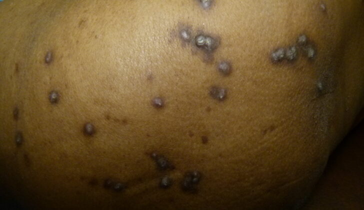
Often, patients with RPC will have bumpy skin growths on the outer parts of their limbs and hands, typically where the skin has been lightly injured. These skin growths are very itchy and can develop into large, belly button-like bumps with a hard center. These bumps come and go throughout a person’s life.
Most instances of RPC resolve on their own and don’t need any treatment.
What Causes Reactive Perforating Collagenosis?
The exact reasons why RPC (a skin condition) happens are still unclear. There are reported cases where RPC appears to be inherited in families, showing patterns of dominant, recessive, or random inheritance. Some experts think that factors like skin injury, genetic risk, changes in small blood vessels, and calcium deposits in the skin might contribute to the development of RPC. In some experiments, people were able to create a typical RPC wound with just a needle scratch.
Risk Factors and Frequency for Reactive Perforating Collagenosis
RPC is a very rare disease that is inherited, with fewer than 50 cases having been reported since its discovery in 1967. However, the form of RPC that adults acquire is more common, affecting about 10% of kidney patients on hemodialysis. This disease affects all races equally, but interestingly, 3 out of the 5 cases of a specific variant of RPC have been found in Asians. RPC is seen in equal amounts in men and women. Usually, the inherited form of RPC is seen in infants or children, while the acquired form usually starts affecting adults from their 30s to their 50s.
Signs and Symptoms of Reactive Perforating Collagenosis
Reactive perforating collagenosis (RPC) is a skin condition marked by small, rough bumps or papules that can appear anywhere on the body. In the inherited version of this condition, which begins in infancy, these bumps tend to show up on the hands, elbows, and knees, often after the skin has been damaged or traumatized in some way. As the condition progresses, these papules can grow into larger, belly button-like lumps with a hard, keratin plug in the center. These lumps usually clear up on their own in about 6 to 8 weeks, often leaving a darker patch of skin or a scar behind.
People with RPC typically report itchiness of the lumps. There can also be a Koebner phenomenon, where skin injury leads to new lumps. The condition usually follows a cycle of flare-ups, followed by periods of healing. A few cases of a larger variant of RPC, where lumps measure between 1 and 10 cm with a central keratin plug, have been observed, including in a 60-year-old woman receiving hemodialysis treatment.
Testing for Reactive Perforating Collagenosis
RPC, or reactive perforating collagenosis, is diagnosed based on certain patterns in the skin (collagen coming out through the skin layers) and specific skin changes such as scaly bumps or indented bumps or nodules. Usually, a procedure called “punch biopsy” is done to collect a small sample of your skin for examination under a microscope. This test helps doctors to confirm the diagnosis of RPC.
There isn’t a genetic test specifically for RPC, but if someone in your family has had it, that could support the fact that you might have it too. Another tool that doctors use to identify RPC is a special device called a dermoscope. This tool can show a yellowish-brown area in the middle of the skin lesion, surrounded by a white ring of varying thickness that indicates accumulation of keratinous (a type of protein) debris, and an outer pink circle with some patterns of tiny blood vessels, which suggests skin inflammation.
Treatment Options for Reactive Perforating Collagenosis
Lesions caused by RPC (Recurrent Papular Crusted Pustular Eruption) often resolve on their own and rarely cause serious issues. The best way to prevent these lesions is by avoiding any harm or injury to the skin, which can increase the risk of developing RPC lesions. If these lesions cause itchiness, they can be managed with skin moisturizers, anti-itch creams, and allergy medicines.
Other treatments such as skin cell softeners, vitamin A creams, oral vitamin A, exposure to special light, freezing with liquid nitrogen, a medicine called methotrexate, and a medicine called allopurinol have been tried but have not shown significant improvement of the lesions.
Clinical trials, which are studies that test the effectiveness of treatments, haven’t been performed specifically for RPC, so suggestions for treatment are based on individual case reports. This means that the treatment can vary greatly from person to person, and can depend on how severe the lesions are.
Most of the time, these lesions go away on their own in about 6 to 8 weeks, leaving changes in skin color (either lighter or darker), scarring, or darkened skin due to inflammation. Unfortunately, it is common for patients to suffer from these lesions on and off throughout their life.
What else can Reactive Perforating Collagenosis be?
When a doctor is trying to determine if a patient has Reactive Perforating Collagenosis (RPC), they may consider other conditions that have similar symptoms. These include:
- Folliculitis: It is characterized by inflamed pustules and bumps where hair grows. Unlike RPC, a biopsy would display inflammation in the hair follicle, not collagen elimination through the skin.
- Prurigo Nodularis: This condition causes more nodular lesions. A biopsy would reveal compact skin overgrowth, irregular skin thickening, and inflammation around blood vessels with thickened collagen in the skin.
- Multiple Keratoacanthomas: These lesions are more like craters than those of RPC. They show a central core of keratin and slight irregular cell growth with a well-differentiated squamous epithelium on a biopsy.
- Dermatofibroma: These generally appear as single, firm, flesh-colored bumps, often on the lower legs. A biopsy would show cells growing in a spindle shape with trapped collagen.
- Arthropod bites: These bites generally do not have dimpled center and tend to show specific inflammatory cells (eosinophilic infiltrates) on a biopsy.
- Other primary perforating skin conditions: These could be Kyrle disease, perforating folliculitis, or elastosis perforans seriginosa.
- Secondary perforating disorders: These could be conditions like lichen nitidus, granuloma annulare, or chromoblastomycosis.
Therefore, a doctor will carefully evaluate the symptoms and possibly perform a biopsy to correctly identify the condition.
What to expect with Reactive Perforating Collagenosis
The inherited form of RPC, a type of skin condition, often carries on into adulthood and can leave some lasting scars. However, for those who acquire the condition later in life, the outlook is generally positive. Some patients even find that itching and skin lesions lessen with treatment for the disease that caused the RPC in the first place.
However, it’s important to note that many people may find that the condition comes back once they stop their treatment.
Possible Complications When Diagnosed with Reactive Perforating Collagenosis
Persistent itchiness can lower a person’s quality of life and increase the risk of a bacterial infection in the skin. Additionally, when the itchy spots heal, they can leave scars and cause changes in skin color, no matter what type of skin the person has.
Effects of Chronic Itching:
- Deterioration of life quality
- Increased risk of bacterial infections on the skin
- Scarring after individual lesions heal
- Skin color alterations in all skin types


