What is Seborrheic Keratosis?
Seborrheic keratosis is a common, harmless skin condition that appears as a variety of shapes and colors. It arises from immature skin cells and looks like a slightly colored patch or a brown to black, textured spot or raised area with a unique “stuck-on” look. This condition mainly occurs in adults and older people. Most people start seeing signs of seborrheic keratosis during their middle adult years.
These spots or patches typically grow slowly in size and number over several years. The condition becomes more common as we get older, and almost everyone who is 60 or older has some. It’s worth noting, however, that it’s a benign condition, meaning it’s not cancerous or harmful.
What Causes Seborrheic Keratosis?
Seborrheic keratosis, a non-cancerous skin condition, happens when immature skin cells called keratinocytes grow excessively. This leads to the formation of well-defined, round or oval, flat spots on the skin, often referred to as macules. These spots generally grow slowly, can thicken over time, and usually don’t go away on their own.
Risk Factors and Frequency for Seborrheic Keratosis
Seborrheic keratosis, a harmless type of skin tumor, affects over 80 million Americans. These growths are typically more common in people over 50 and tend to become more noticeable as people get older. However, they can also be found in younger adults. Both males and females can develop seborrheic keratosis without any significant difference in occurrence. But, it’s important to note that these skin growths appear more commonly in those with lighter skin tones.
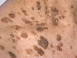
Signs and Symptoms of Seborrheic Keratosis
Seborrheic keratoses are skin growths that have a distinct “stuck-on” appearance due to their waxy and bumpy texture. The color can vary from light to dark brown, yellow, or grey, and they can either occur as a single lesion or as many as hundreds. Commonly, these can be found anywhere except for the palms, soles, and mucous membranes. Frequent sites include the face, scalp, trunk, under the breasts, arms, and legs. While usually not causing any symptoms, if they become inflamed or irritated, they can cause itching or mild pain. Although they’re often harmless and slow-growing, a sudden growth or burst of multiple its lesions can be a sign of underlying issues, such as gastrointestinal or lung cancer.
There are specific types of seborrheic keratoses, including:
- Stucco keratoses, which are small, white bumps often appearing on the feet and ankles. There’s debate over whether these are a form of seborrheic keratosis or not.
- Dermatosis papulosa nigra. This is a variant that tends to develop in darker-skinned individuals. It shows up as brown to black bumps, mostly on the face and neck.
- The Leser-Trélat sign, characterized by a sudden onset of multiple seborrheic keratoses as well as skin tags. This has been linked to various cancers.
- The pseudo-Leser-Trélat sign, characterized by inflammation in existing seborrheic warts after chemotherapy.
Testing for Seborrheic Keratosis
Seborrheic keratosis is a skin condition that is usually identified by doctors through a simple observation because of its distinct look. It appears as a raised, brownish or blackish skin bump that looks like it’s stuck to the skin and has a scaly texture.
Doctors can also use a device called a dermoscope to get a closer look at the skin bump. This device can show particular features that suggest the bump is a seborrheic keratosis, such as openings that look like clogged pores, tiny fluid-filled sacs, skin folds, and certain types of blood vessels. While these skin bumps can look a lot like some types of skin cancer, they usually lack certain patterns of color and shape that appear in cancerous bumps.
Even though seborrheic keratosis is usually identified by sight alone, there might be times when it’s not that clear. In cases where the doctors aren’t sure, they might need to take a small sample of the bump to examine it under a microscope to ensure that it’s not a skin cancer such as melanoma, squamous cell carcinoma, or basal cell carcinoma.
Seborrheic keratosis is generally a slow-growing skin condition that can gradually get bigger and thicker over time. There have been instances where another type of skin growth has appeared next to or even inside an existing seborrheic keratosis. If a patient has lots of these skin bumps and there is a chance they might be cancerous, a skin specialist, known as a dermatologist, should be consulted.
If the diagnosis is uncertain or there’s concern that the skin bump might be cancerous – if it’s got a sore that won’t heal, quickly gets bigger or is very large, for instance – then a skin biopsy is advised to confirm the diagnosis. There are certain situations, like if a patient suddenly gets a lot of seborrheic keratoses at the same time, that could suggest an underlying cancer such as gastrointestinal tract cancer, leukemia, or lymphoma. This is known as the Leser-Treélat sign. For these individuals, it’s very important that they get a comprehensive medical assessment, cancer screening if appropriate for their age, and any necessary lab tests based on their health history and risk factors. Other reasons for a sudden outbreak of multiple seborrheic keratoses could be chemotherapy treatment or a skin inflammation condition called eczema.
Treatment Options for Seborrheic Keratosis
Seborrheic keratosis is a common, harmless skin growth often found in adults. These growths often don’t require treatment except in a few cases. For instance, if the growths change rapidly, bleed, or create discomfort, it might be necessary to remove them because they have a higher risk of becoming malignant, or cancerous. The method of removal depends on several factors such as the size and thickness of the growth, the skin type of the patient, the chances the growth might be cancerous, and the doctor’s experience.
There are several procedures available to remove seborrheic keratosis:
1. Cryotherapy: This is the commonly used treatment and is generally well-tolerated by patients. In this method, the doctor uses liquid nitrogen or carbon dioxide to rapidly freeze or thaw the growth, which leads to the death of the growth’s cells. While generally effective, this procedure can cause some temporary skin redness, pain, and blister formation. In some cases, it may also cause longer-term changes in skin color in the treated area.
2. Electrodesiccation and curettage: A process is another option to remove superficial, skin-deep growths. This procedure usually requires local anesthetics and can be done in the doctor’s office. The doctor first scrapes off the growth with a special instrument called a curette, and then uses an electric instrument to destroy the remaining tissue.
3. Ablative laser treatment: In this procedure, the doctor uses high-energy light (laser) to vaporize the growth. There are several types of lasers that can be used, and this approach has shown promise in some studies, but there’s a need for more research to establish its efficacy.
4. Shave biopsy: With this technique, the doctor numbs your skin and then uses a razor blade to shave off growths that have not penetrated deeply into the skin. This method allows the doctor to study the tissue under a microscope to confirm that the growth is indeed a seborrheic keratosis.
In addition to these procedures, various types of topical agents are being studied for treating seborrheic keratosis. These are gels and creams that you apply to the skin. Early studies suggest some of these might help, but more work is needed to prove their effectiveness. The U.S. Food and Drug Administration recently approved a solution of 40% hydrogen peroxide for treating seborrheic keratosis. Trials have found it to be generally well-tolerated and effective.
What else can Seborrheic Keratosis be?
When doctors are trying to diagnose seborrheic keratosis (a common type of harmless skin growth that resembles a wart), they have to consider a range of other conditions that have similar features. These include:
- Malignant melanoma (a type of skin cancer)
- Actinic keratosis (rough, scaly patches caused by sun damage)
- Lentigo maligna (a type of skin cancer that usually occurs in older people)
- Melanocytic nevus (moles)
- Squamous cell carcinoma (a type of skin cancer)
- Pigmented basal cell carcinoma (a type of skin cancer).
If a patient has lots of seborrheic keratosis growths, it can be harder for the doctor to diagnose and examine them. This is because there’s a higher risk of missing any cancerous growths that could be mixed in.
Melanoma, for example, is an especially tricky condition to separate from seborrheic keratosis. That’s because there are some rare instances when melanoma can look like seborrheic keratosis. Usually, though, melanoma and most seborrheic keratoses look and behave differently. Melanoma tends to be flat and spreading, while most seborrheic keratoses are raised and rough. If a growth is difficult to diagnose, the doctors may need to carry out extra tests and examinations to make sure they’ve got it right.
Seborrheic keratosis can also look like pigmented actinic keratosis, pigmented basal cell carcinoma, or cutaneous squamous cell carcinoma, especially when they appear on the face or other areas that get a lot of sun exposure. However, these conditions don’t have the characteristic ‘stuck-on’ appearance of seborrheic keratosis.
Other conditions that need to be considered in the diagnosis are epidermal nevus (a kind of birthmark), acanthosis nigricans (a skin condition causing darkened, thickened patches), and viral warts. For example, rough seborrheic keratosis can look a lot like verruca vulgaris (common warts), especially if they’re in the genital or perineal region. In those situations, doctors might need to test for human papillomavirus (HPV) or take a biopsy to figure out what’s going on.
What to expect with Seborrheic Keratosis
Seborrheic keratoses are non-cancerous growths and generally have a very good outlook. Most of the time, they are treated for appearance-related reasons. Various treatment methods can successfully remove these growths with very few side effects or complications.
If a person has many seborrheic keratoses, it’s crucial to examine them closely. This is because the large number of growths could hide associated cancerous growths. There’s a condition known as Leser-Trelat, which indicates internal cancer and usually suggests a worse outlook.
Possible Complications When Diagnosed with Seborrheic Keratosis
Seborrheic keratoses are harmless skin growths that are very common. They grow slowly and might get thicker as time goes on. Depending on where they are on your body, they could get irritated and become painful or uncomfortable. If you have a lot of them, it can be hard to spot other harmful skin growths.
There is a medical case where a seborrheic keratosis turned into another type of skin growth called bowenoid actinic keratosis. This was in a person who had rheumatoid arthritis and was taking multiple medicines to lower their immune system’s response. Three specific changes in the tissue’s appearance caused the transformation.
Preventing Seborrheic Keratosis
Preventing and educating patients are key in managing seborrheic keratosis, a common harmless skin condition. Doctors have a vital role in helping patients understand that seborrheic keratosis is not dangerous or cancerous. They stress the need for regular skin checks to identify any changes early on. Education for patients should include tips on how to protect their skin from the sun, which can help avoid new spots and keep current ones from worsening.
Doctors should also explain the different treatment options for seborrheic keratosis, like cryotherapy (freezing off the growths), curettage (scraping off the growths), and creams or lotions that can be applied to the skin. Knowing about these treatments allows patients to make informed choices about their healthcare.
By promoting understanding and encouraging good skin care practices, doctors can help reduce patient worry, aid in early detection, and ultimately improve the health of people with seborrheic keratosis.


