What is Subungual Melanoma?
Longitudinal melanonychia is a condition where there are dark streaks that run down the nails. It comes in three different forms: lentigo, nail matrix nevus, and subungual melanoma. The first two types are harmless, but subungual melanoma is a type of skin cancer that originates from the structures within the nail.
Subungual melanoma is typically a form of another skin cancer called acral lentiginous melanoma, which usually appears on the palms of the hands and soles of the feet. It was first discovered in 1834 and was later detailed in 1886, when a specific sign called the “Hutchinson sign” was described, indicating the spread of pigment beyond the nail onto the adjacent skin.
Although subungual melanoma is rare, it accounts for up to 3 percent of all melanoma skin cancers in people with light skin, and up to 30 percent in people with dark skin. Most commonly, it appears as a dark black, vertical band on a single nail, often wider than 3 millimeters. The edges of the band may be irregular, and the nail itself can appear damaged or distorted.
What Causes Subungual Melanoma?
Malignant melanoma is a type of cancer that starts from the pigment-producing cells called melanocytes. When it occurs in the nail matrix, the region where your nails start to grow, it’s known as subungual melanoma. Unlike malignant melanoma that occurs on the skin and is often related to sun exposure, subungual melanoma doesn’t seem to be linked with sunlight.
Risk Factors and Frequency for Subungual Melanoma
Subungual melanomas, a type of skin cancer that affects the nail area, are quite rare, making up between 0.7% to 3.5% of all malignant melanomas globally. Regardless of race, people can get these melanomas. Interestingly, due to their natural skin protection against harmful sun rays, people of African, Asian, and Hispanic descent commonly get this variant of melanoma. Among these groups, this skin cancer forms 75% of all melanomas in Africans, 25% in Chinese, and 10% in Japanese.
People in their sixties and seventies are most likely to get it, with women getting it a bit earlier than men. The thumb and great toe are the fingers generally affected, with 75% to 90% of subungual melanomas cases happening there.
Signs and Symptoms of Subungual Melanoma
Subungual melanoma is a type of skin cancer that shows up as a brownish-black mark on the nail bed. It may appear in the form of a coloring stripe or uneven coloration. Over time, this discoloration can lead to thickening or even damage of the nail, accompanied by pain and inflammation. Sometimes, there can be coloration around the nail (a sign called Hutchinson sign), which often indicates melanoma.
However, the Hutchinson sign isn’t always proof of melanoma. The same kind of coloration can occur due to other conditions such as nail freckles or marks, melanonychia (a condition common among individuals with dark skin tones), Laugier-Hunziker syndrome, or Peutz-Jeghers syndrome. When the coloration happens due to these conditions, it’s referred to as the “pseudo-Hutchinson sign”.
It’s important to remember incidents of injury when diagnosing subungual melanoma as it could be mistaken for a blood clot under the nail (subungual hematoma). An infection resulting in pigmented fungal nail conditions should also be ruled out.
To evaluate the risk of a discolored nail lesion being a melanoma, the ABCDEF guidelines suggested by Levit et al. can be used:
- Age: Individuals between 50 to 70 years, or of African, Japanese, Chinese, and Native American descent
- Brown-black band: Greater than 3 mm with an irregular outline
- Change: Alteration in size and growth speed
- Digit: Particularly affects the thumb, big toe, or index finger
- Extension: Discoloration spreading into the skin around the nail (Hutchinson sign)
- Family history: Past family cases of melanoma
Testing for Subungual Melanoma
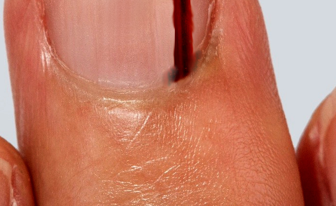
If a doctor suspects there may be a problem with a patient’s nail bed (the skin beneath the nail), they’ll need to perform a procedure known as a full-thickness biopsy to help diagnose the issue. Research shows that it’s important to get a good quality biopsy to ensure the initial examination is accurate. After this, the doctor can create a plan for dealing with the issue.
Here’s how a nail bed biopsy works: To prevent pain, the doctor uses a digital nerve block, which is a type of anesthesia. Although a tourniquet (a device used to apply pressure) can help control bleeding, it’s important not to drain all the blood from the nail bed in case there’s a malignant (cancerous) growth. The doctor will then carefully remove the nail so they can see the germinal matrix (the part of the nail where new nail cells are produced). To get a good view, the doctor may make two small cuts at 45 degrees against the eponychial fold (the skin at the base of the nail). Then, they’ll remove a small piece of the pigmented (colored) area to send to the lab for testing — this is the biopsy. If possible, the doctor will try to remove the entire lesion (abnormal growth). Afterwards, the doctor will close the wound with a dissolvable suture (stitch) and replace the nail.
Once the biopsy has been analyzed, a treatment plan can be made based on the results. The lab will provide information on what type of malignant melanoma (a type of skin cancer), if any, is present, how deep it is (the Breslow thickness), and other details about its appearance under the microscope. This information is crucial to determine the best way to treat the nail bed issue.
Treatment Options for Subungual Melanoma
When it comes to treating melanoma, or skin cancer, there’s a team made up of various specialists that will review your case. They’ll use the information collected about your tumor to determine the best course of action in terms of testing and treatment. Typically, the go-to treatment for any type of melanoma is removing it via surgery.
The first part of this process is a biopsy, which involves removing a small piece of the tumor for testing. The test results will give doctors an idea about how deep the tumor is. After that, using established guidelines, the doctors will decide how much additional tissue needs to be removed around the tumor. This is the standard procedure whether the melanoma is under the nail (subungual) or elsewhere.
Besides this, doctors will examine lymph nodes, which is another way cancer can spread. If they’re found to be suspicious during the physical exam, doctors will suggest further imaging tests, and possibly a biopsy. Depending on the results, they might consider removing these lymph nodes.
For cases where the melanoma is between 1 to 4 mm thick and no enlarged lymph nodes are identified, patients may be offered what’s called a sentinel lymph node biopsy. This is a procedure to check if the melanoma has started to spread.
If the melanoma is thicker than 4 mm, doctors may recommend scans such as CT-PET or brain MRI to check the extent of the disease.
The pathway for treating melanoma keeps evolving and is quite complex. For instance, there’s consensus that a biopsy that removes the entire growth is the best way to diagnose melanoma. However, there’s no hard evidence that using other biopsy types impacts how long patients survive or how often the cancer comes back. According to the National Comprehensive Cancer Network guidelines, one should remove additional tissue around the tumor based on the Breslow depth, which is a measure of thickness of the tumor. For melanomas of medium thickness (1.0 to 4.0 mm), doctors typically recommend a sentinel lymph node biopsy. The same process might be considered for melanomas as thin as 0.75 to 1.0 mm but carry high risks due to characteristics like ulceration or an increased number of cells dividing.
What else can Subungual Melanoma be?
Two conditions often mistaken for subungual melanoma, a type of skin cancer that develops under the nail, are striate melanonychia and onychomycosis. Striate melanonychia is a condition where melanin, the pigment that gives color to our skin, hair, and eyes, accumulates in the nail – typically not as a single pigmented streak though. Onychomycosis, on the other hand, is a painless fungal nail infection that causes changes in the nail’s color and texture.
Aside from these two conditions, other potential diagnoses that must be considered are subungual hematoma, which is a painful bruise under the nail that usually occurs after an injury, and junctional naevus, which is a benign skin lesion that doesn’t have a streaked pattern like that often seen in subungual melanoma.
When a doctor is evaluating a patient for possible subungual melanoma, it’s important that they also consider and rule out subungual hematoma and nail bed infections.
Surgical Treatment of Subungual Melanoma
Subungual melanoma is a rare type of skin cancer that develops under the nail. Medical professionals have debated the best way to treat this disease for many years. In the past, the only acceptable treatment was amputating the affected finger or toe. A study in 1965 observed that all patients who had amputations at the joint closest to the nail didn’t live for five years beyond their treatment. Despite this study’s significant findings, it’s important to note that the authors didn’t report how deep the cancer was in the patients, and half of the patients already had cancer that had spread or come back at the time of their diagnosis.
Since then, doctors have started to reconsider such aggressive treatment methods. The shift in the medical community has prompted research into less drastic surgical options, with the goal of preserving the length of the affected finger or toe and ensuring the cancer is completely removed. A review of available literature in 2014 found that for lower-level melanomas (a type of skin cancer), removing the cancer without amputating the finger or toe is a sensible approach. Similarly, for deeper, higher-level melanomas, amputation could be performed at a less severe level than previously recommended.
However, the available data supporting these less aggressive treatment options isn’t of high quality. There aren’t any results from strong scientific studies, like randomized controlled trials, and most of the data has been collected after the fact. As a result, there isn’t a one-size-fits-all solution, and the surgical approach should be decided on a patient-by-patient basis.
To help medical professionals make these decisions, here are some guidelines based on how deep the melanoma is:
- For melanoma in situ (meaning the cancer cells are only in the outermost layer of the skin) that is 0.5 cm to 1 cm deep: surgical excision of 1 cm around the melanoma.
- For invasive melanoma less than 1 mm deep: surgical excision of 1 cm around the melanoma.
- For melanoma 1 to 2 mm deep: surgical excision of 1-2 cm around the melanoma.
- For melanoma greater than 2 mm deep: surgical excision of 2 cm around the melanoma.
These guidelines need to be checked against the patient’s everyday functionality.
If the patient has cancer that can be felt in the lymph nodes, they should receive surgery to remove them. Guidance from the UK suggests that patients with melanoma that is 1 to 4 mm deep should be offered a procedure to test the cancer status of the closest lymph node. If this test shows signs of spread, then all the lymph nodes in the area should be removed.
What to expect with Subungual Melanoma
Some studies suggest that subungual melanoma, a type of skin cancer that develops under the nail, might have a worse prognosis than other skin melanomas. Importantly, this is often because people discover or seek treatment for this condition late. Nevertheless, if detected and treated early, the prognosis for subungual melanoma should be about the same as for other skin melanomas.
Here are some recent survival rate statistics for skin melanoma, provided by the American Cancer Society:
- Stage IA: About 97% of patients are expected to live for at least 5 years, and about 95% for 10 years.
- Stage IB: About 92% of patients are expected to live for at least 5 years, and about 86% for 10 years.
- Stage IIA: About 81% of patients are expected to live for at least 5 years, and about 67% for 10 years.
- Stage IIB: About 70% of patients are expected to live for at least 5 years, and about 57% for 10 years.
- Stage IIC: About 53% of patients are expected to live for at least 5 years, and about 40% for 10 years.
- Stage IIIA: About 78% of patients are expected to live for at least 5 years, and about 68% for 10 years.
- Stage IIIB: About 59% of patients are expected to live for at least 5 years, and about 43% for 10 years.
- Stage IIIC: About 40% of patients are expected to live for at least 5 years, and about 24% for 10 years.
- Stage IV: About 15% to 20% of patients are expected to live for at least 5 years, and about 10% to 15% for 10 years.
For Stage IV, the survival rate can be better if the cancer has only spread to distant parts of the skin or to lymph nodes far from the original cancer, and if the patient’s blood level of a substance called lactate dehydrogenase (LDH) is normal.
Possible Complications When Diagnosed with Subungual Melanoma
After an operation, some people might experience a condition called nail dystrophy. In simple terms, nail dystrophy alters the natural structure and appearance of the nails. Also, all patients tend to witness some form of aesthetic changes post-surgery.
Common Side Effects:
- Nail dystrophy
- Cosmetic deformities
Recovery from Subungual Melanoma
Patients who choose not to have a biopsy need to be watched carefully by their healthcare professionals.
Preventing Subungual Melanoma
It’s important for patients to realize that subungual melanoma, a type of skin cancer that forms in the tissues of the nail bed, is not linked with exposure to the sun. Maintaining good hygiene of the hands and feet may be beneficial. Furthermore, regularly checking their nails and the skin beneath them for any new spots or return of the abnormal growth can help in early detection and treatment.


