What is Tinea Barbae?
Dermatophytes are types of fungi that attack tissues containing a protein known as keratin, found in skin, hair and nails. An infection caused by these fungi is known as dermatophytosis, commonly called a “tinea” infection. These fungi primarily feed on keratin, which is why they don’t infect the mucosal surfaces that lack this protein. Dermatophyte infections can occur on any surface of your body that has keratin, from your scalp down to your toes. These infections are named based on the body part they affect.
One rare type of dermatophyte infection, Tinea barbae, largely affects the beard and mustache area, including the hair and hair follicles. First identified by a researcher named Gruby in 1842, this condition was noticed as fungus forming a protective layer around hair in the beard area. Gruby called this fungus “mentagrophyte”, meaning “plant of the chin”. This infection often inflames the hair follicles, leading it to also be referred to as tinea sycosis. It has additionally been known as barber’s itch or beard ringworm, on account of its historical association with unsanitary razors used by barbers.
What Causes Tinea Barbae?
People get dermatophyte infections, a type of fungal infection, by coming into direct contact with infected people or animals, or through soil. This type of infection includes a specific condition known as tinea barbae, which only comes from contact with infected animals or people.
Several different types of fungi can cause these infections, but the most common are Trichophyton verrucosum, Trichophyton mentagrophytes, and Trichophyton rubrum. There are a few other, less common types of fungus that can also cause these infections.
Until the year 2000, most reported cases of tinea barbae were traced back to animals, particularly farm and domestic animals like dairy cows, sheep, pigs, horses, dogs, and cats. However, more recent reports show that the Trichophyton rubrum fungus, which passes from person to person, is becoming a frequent cause of these infections. According to some cases recorded in Portugal and Mexico, tinea barbae can sometimes be transmitted from one person to another, but this is rare.
There have even been a few cases where patients with a foot infection, known as tinea pedis, were able to transmit the infection to their own beards. This process is known as ‘autoinoculation’.
Risk Factors and Frequency for Tinea Barbae
Tinea barbae is a specific type of skin fungus infection that’s not very common, despite the fact that skin fungus infections in general are quite common around the world. It was first recorded in 1842, and since then, there have been only around 150 cases reported until 1990. Most reports after 1990 are isolated incidents or small groups of cases.
Because it’s quite rare, it’s hard to say how frequently it occurs or how many people might get it in their lifetime. The fungus that causes it can be found all over the world, and reported cases of tinea barbae have come from various countries. Tinea barbae affects the hair and hair follicles in the beard and mustache area, so it’s only seen in boys who are old enough to grow facial hair and in men. However, there has been an extremely rare case reported in a woman from Portugal who had a significant amount of facial hair.

Signs and Symptoms of Tinea Barbae
Tinea barbae is a relatively uncommon skin infection caused primarily by a type of fungus known as zoophilic fungi. Its diagnosis can be helped by understanding the disease well and looking at the patient’s history, including their occupation, contact with pets or farm animals, and conditions such as diabetes mellitus, past injuries, use of steroids, and immunosuppressant therapy. All these factors might impact the local defense mechanisms against the fungus. It’s also important to check if the patient or their close contacts have a history of similar fungal infections like tinea capitis, tinea pedis, etc.
Tinea barbae has two main forms, one that causes inflammation and another that doesn’t. The former is linked to zoophilic dermatophytosis, a group of fungus that primarily infects animals. The latter is associated with anthropophilic dermatophytosis, the fungi that typically infect humans.
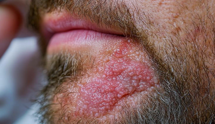
- The inflammatory form of tinea barbae causes a distinct lesion known as a kerion, which is a reddish, tender, boggy, often weeping lump or patch of skin with pustules and draining sinuses. The hair in these affected parts loses its shine, becomes brittle, and is easily plucked. The kerion can be solitary or multiple but usually affects just one side. Symptoms may extend beyond the skin and include fever and fatigue. Rarely, these lesions can become super-infected with skin bacteria and cause swollen lymph nodes.
- Non-inflammatory tinea barbae is a form of skin infection that causes itching. It presents as a diffusely red, scaly patch with pustules nearby hair follicles. Occasionally, its customarily flat, scaly patches with central clearing can appear similar to facial ringworm, characterized by a ring-shaped border consisting of vesicles and pustules.
Testing for Tinea Barbae
Tinea barbae, a fungal infection that affects the hair and skin, is mainly diagnosed by closely examining its symptoms and understanding its typical traits. Understanding the patient’s history and conducting a thorough physical examination will also contribute to an early diagnosis. Because the common treatment for tinea barbae requires the use of antifungal medication over a long period of time – which could lead to side effects – confirming the diagnosis is essential.
Doctors often confirm the diagnosis by either examining skin scrapings from the infected area under a microscope or by using a special ultraviolet light called a ‘Wood’s lamp’ to inspect the affected area. However, the latter method may have limited usefulness since only a certain type of dermatophyte (fungus) that causes tinea barbae, known as Microsporum, will resonate under the light.
Direct microscopic examination involves obtaining scales and skin scrapings from the lesion area using a scalpel or a moist cotton tip. This sample is placed onto a slide and mixed with a chemical solution (potassium hydroxide) to dissolve the skin cells. Sometimes the slide is gently warmed or another chemical, dimethyl sulfoxide, is added to speed up this process. Fungal stains may be used to make it easier to see the fungus under the microscope. The result will show fungal filaments, like tiny rod-shaped branches, and spores, known as arthroconidia, which confirms the diagnosis.
In some cases, a fungal culture can be done, where the skin scrapings and hair samples are placed into a special growth medium to cultivate the fungus. It typically takes one to two weeks for the fungus to grow, and if no growth is seen for 21 days, the culture is considered negative. Although fungal cultures can take time and be more costly, they can be more precise in identifying the fungus compared to direct microscopy. This method can assist in tailoring the treatment process but isn’t always required before antifungal treatment can begin.
For more complex cases where results aren’t conclusive with previous tests, a skin biopsy can be performed and examined under a microscope. A pathologist looks for signs like fungal structures, inflammation changes, and the presence of folliculitis, perifolliculitis, and microabscesses. More advanced DNA-based testing techniques exist, but these are currently expensive and used mostly in research centers.
Treatment Options for Tinea Barbae
Tinea barbae, a skin infection affecting bearded areas of the face and neck, is primarily treated with oral antifungal medicines. While topical treatments can be applied to the skin, these are typically used alongside oral medications, not as the main treatment.
Effective oral antifungal medicines for tinea barbae include terbinafine, azoles, and griseofulvin. In the past, griseofulvin was often the preferred choice, but it’s now used less due to its side effects, the potential for drug resistance, the requirement for long-term treatment, and a higher likelihood of the infection returning due to the medicine quickly leaving the skin.
Previous treatment plans with griseofulvin often lasted for 12 weeks. However, more recent reports have shown that tinea barbae can be effectively treated with terbinafine and azoles within 4 to 6 weeks. Even once the skin lesions are resolved, some experts suggest continuing treatment for an additional 2 to 3 weeks.
You should be aware that common side effects for terbinafine and azoles can include nausea, abdominal pain, diarrhea, rash, elevated enzyme levels in the liver, and visual disturbances. So, if you are prescribed these medicines, periodic checks of your liver, kidney, and blood functions may be recommended. You should also be mindful of potential interactions between these antifungal medications and other drugs that are broken down in the liver.
If a secondary bacterial infection is suspected, oral or systemic antibiotics may be required. The use of steroid treatments and other topical agents, such as selenium sulfide, is less clear and is typically decided on a case-by-case basis. In rare instances, surgery could be an option, though draining infections through an incision is not usually recommended.
What else can Tinea Barbae be?
Tinea barbae, an uncommon condition that affects the beard and mustache area, is often mistaken for more frequently occurring conditions. These might include bacterial folliculitis, perioral dermatitis, pseudofolliculitis barbae, contact dermatitis, herpes simplex, sporotrichosis, acne vulgaris, acne rosacea, and even rare cases of neoplasm. Understanding the distinguishing characteristics of these conditions can help with earlier diagnosis of tinea barbae.
For instance, bacterial folliculitis caused by Staphylococcus species or other bacteria can be differentiated from tinea barbae. This, if systemic symptoms like fever and malaise are present and there is a prevalence of regional lymphadenopathy. Plus, removing hair is painless in the case of tinea barbae, unlike with sycosis barbae. Pseudofolliculitis barbae, caused by shaving irritation, is bilateral and diffuse, contrasting with tinea barbae’s presentation. And, candidal infections, unlike tinea barbae, affect adjacent mucosal surfaces.
What to expect with Tinea Barbae
Tinea barbae, a rare type of skin fungal infection, hasn’t been widely reported with only a few hundred known cases in the medical literature. Many of these recorded instances of tinea barbae have been individual accounts or small collections of cases.
Despite the limited amount of data on this condition, it is encouraging to note that nearly all reported cases have shown a positive response to treatment with oral antifungal medication, resulting in complete healing.
Possible Complications When Diagnosed with Tinea Barbae
Other than health issues and the costs related to the treatment, there have not been any major complications reported in medical studies or documents.
Preventing Tinea Barbae
Tinea barbae is a contagious skin infection that you can catch from humans or animals, either through direct touch or indirect contact like using the same items. If you’re diagnosed with tinea barbae, it’s important to figure out how and where you got it. It’s suggested to check yourself and your family members for similar skin infections like tinea capitis (a fungal infection of the scalp), tinea pedis (known commonly as athlete’s foot), and onychomycosis (a fungal infection in the nails).
It’s also a good idea to have any pets or farm animals checked for similar infections by a veterinarian. This can prevent the infection from coming back. To avoid spreading the infection among family members, avoid sharing personal items like combs, brushes, and hats.


